1791
Novel application of the reversed gradient method in Diffusion Weighted-MRI for tumor response assessment in head and neck squamous cell carcinoma patient undergoing radiation therapy.David Aramburu Nuñez1,2,3, Jose Luis del Olmo Claudio3, Silvia Reigosa Montes3, Antonio López Medina3, Moises Mera Iglesias4, Francisco Salvador Gómez 3, Íñigo Nieto5, Alfonso Calzado2, Amita Shukla-Dave6, and Victor M Muñoz5
1Medical Physics, Memorial Sloan-Kettering Cancer Center, NEW YORK, NY, United States, 2Department of Radiology, Complutense University, MADRID, Spain, 3Department of Medical Physics and Radiological protection, Galaria - Hospital do Meixoeiro – Complexo Hospitalario Universitario de Vigo, VIGO, Spain, 4Medical Physics, Oncoserv, Santiago de los Caballeros – Dominican Republic, Dominican Republic, 5Department of Radiation Oncology, Galaria - Hospital do Meixoeiro – Complexo Hospitalario Universitario de Vigo, Spain, 6Departments of Medical Physics & Radiology, Memorial Sloan-Kettering Cancer Center, NY, United States
Synopsis
Reversed gradient method can reduce geometric distortion leading to
Purpose
The most challenging issue in radiation therapy is the non-invasive, accurate assessment of tumor response during treatment. Apparent diffusion coefficient (ADC)1, derived from DW-MRI has shown promise in tumor characterization and assessment of treatment response2. However, geometric distortions associated with the DW-MRI data collected in clinical setting needs to be corrected prior to the calculation of ADC. One of the options for this correction is to use reversed gradient method3,4. This pilot study investigates the use of the reversed gradient correction method in DW-MRI forMethods
MRI data acquisition: We developed a new phantom for distortion assessment (DA) in DW-MRI (Figure 1). It consists of a 17 x 17 x 17 cm³ polyethylene HD 500 cube, which contains 10 rows and 5 columns of cylindrical tubes (diameter: 14 mm) filled with water and capped at the ends. After phantom studies were performed at room temperature, three head and neck squamous cell carcinoma patients (stage IV) were enrolled for in vivo feasibility study and was approved by local institutional review board. The patients had 4 MRIs (1pre-, 2Results
Phantom study: No significant difference was observed in ADC values of water derived from the raw ((2.13 ± 0.19) × 10-3 mm²/s) and corrected ((2.19 ± 0.05) × 10-3 mm² /s) DW-MRI data at room temperature. The mutual information metric values reflect their dependence on the b-value (13% for b=0, 23% for b=600 and 36% for b=1000s/mm2) [Table 1]. PatientsDiscussion
ADC has shown promise forConclusion
The reverse gradient method may find wider applicability after validation in a larger cancer patient population for tumor response assessment which needsAcknowledgements
The National Health Institute of Spain is supporting this work by the ISCIII Grant PI11/02035 and DTS14/00188, and BIOCAPS project (FP7/REGPOT-2012-2013.1 under grant agreement n° 316265) that also partially supported this research.References
1. Stejskal, E. O. and Tanner, J. E., "Spin diffusion measurements: spin echoes in the presence of aFigures
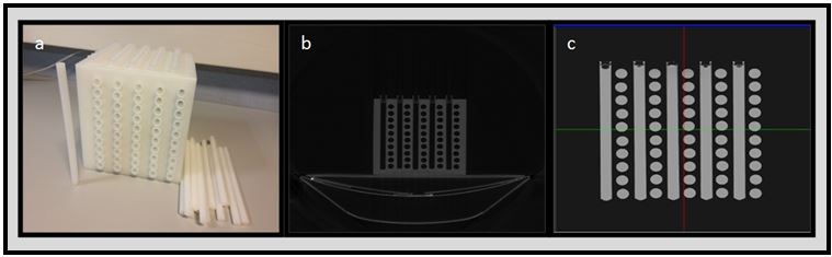
Figure.1.
a. Phantom-DA, specifically designed
for distortion assessment in DW-MRI. b.
CT Axial image of empty Phantom-DA, c.
T2w MRI Axial image of Phantom-DA, Cylindrical hollows are filled with water.
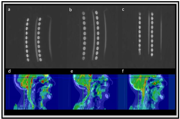
Figure.2.
The upper figure shows the DW-MRI with b=0 for the Phantom-DA: a. Phase
encoding direction: Antero-Posterior, fat shift anterior (AP); b. Phase
encoding direction: Antero-Posterior, fat shift posterior (PA); c. Corrected by
the reversed gradient method. The lower figure shows the registration of CT and
DW-MRI with b=0 for a patient: d. Phase
encoding direction: Antero-Posterior, fat shift anterior (AP); e. Phase
encoding direction: Antero-Posterior, fat shift posterior (PA); f. Corrected by
the reversed gradient method.
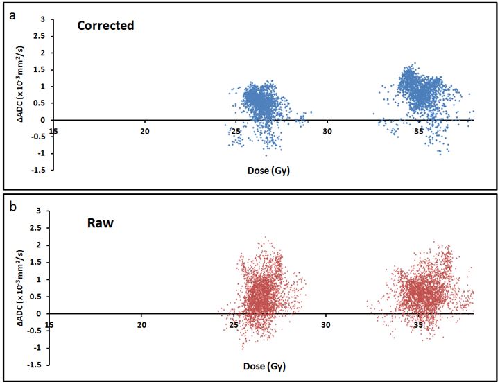
Figure.3.
ΔADC (10-3 mm2/s) vs Dose (Gy) in the tumor. Each point
represents the voxel ΔADC values extracted from a. DW-MRI RAW, b. DW-MRI
corrected at the 12th and 16th fractions of Treatment.
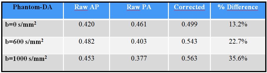
Table 1.
Mutual Information values for the registration of T2w MRI and DW-MRI with
different b-values of the phantom-DA. % difference is calculated with respect
to averaged mutual information of raw studies, considering both gradients. Raw
AP and raw PA correspond to the reversed gradients applied for phase encoding
direction in the AP dimension and different fat shifts.
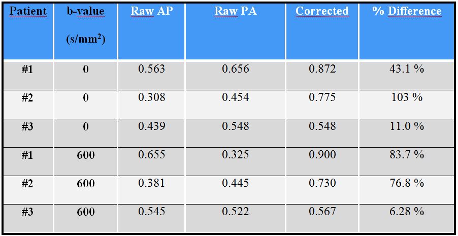
Table 2.
- Mutual information values for different DW-MRI of the three patients with
different b-values and T2w MRI. % difference is calculated with respect to
averaged mutual information of raw studies, considering both gradients. Raw AP
and raw PA correspond to reversed gradients applied for phase encoding
direction in the AP dimension and different fat shifts.