1776

Characteristic analysis of in vivo and ex vivo rat brains by 7.0 T DTI MR
Chunhua Wang1, Li Song, Ruzhi Zhang, and Fabao Gao

1Department of Radiology, West China Hospital, Chengdu, People's Republic of China

Synopsis

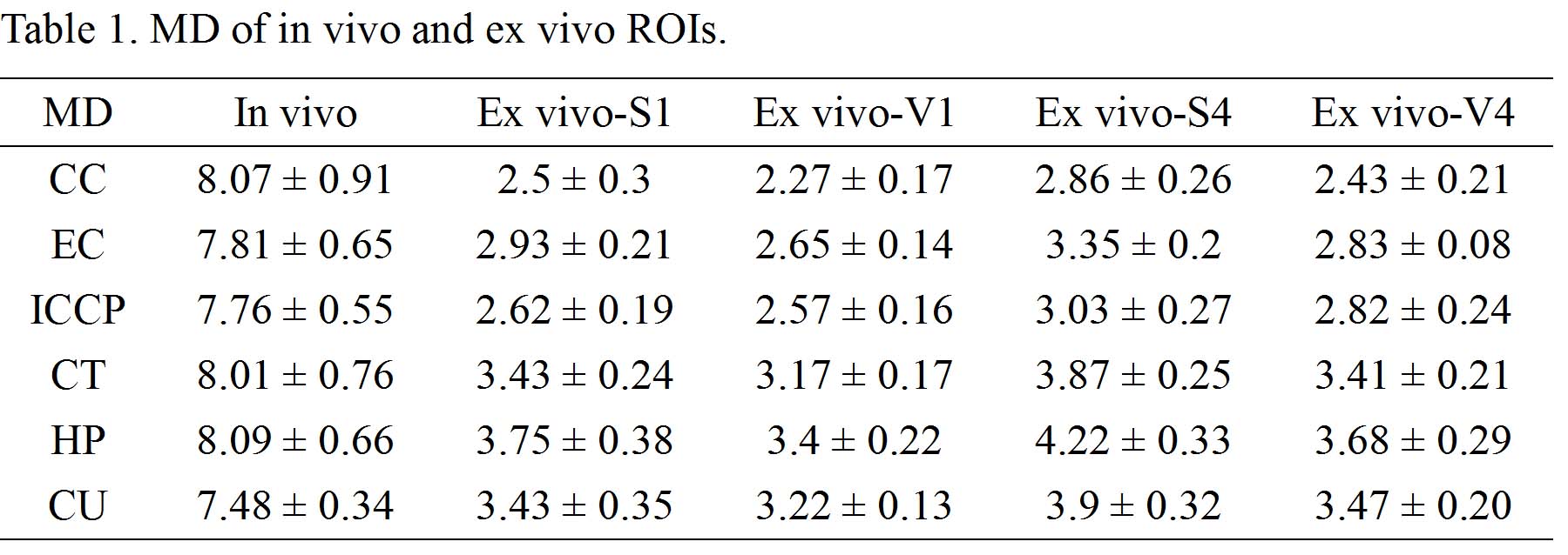
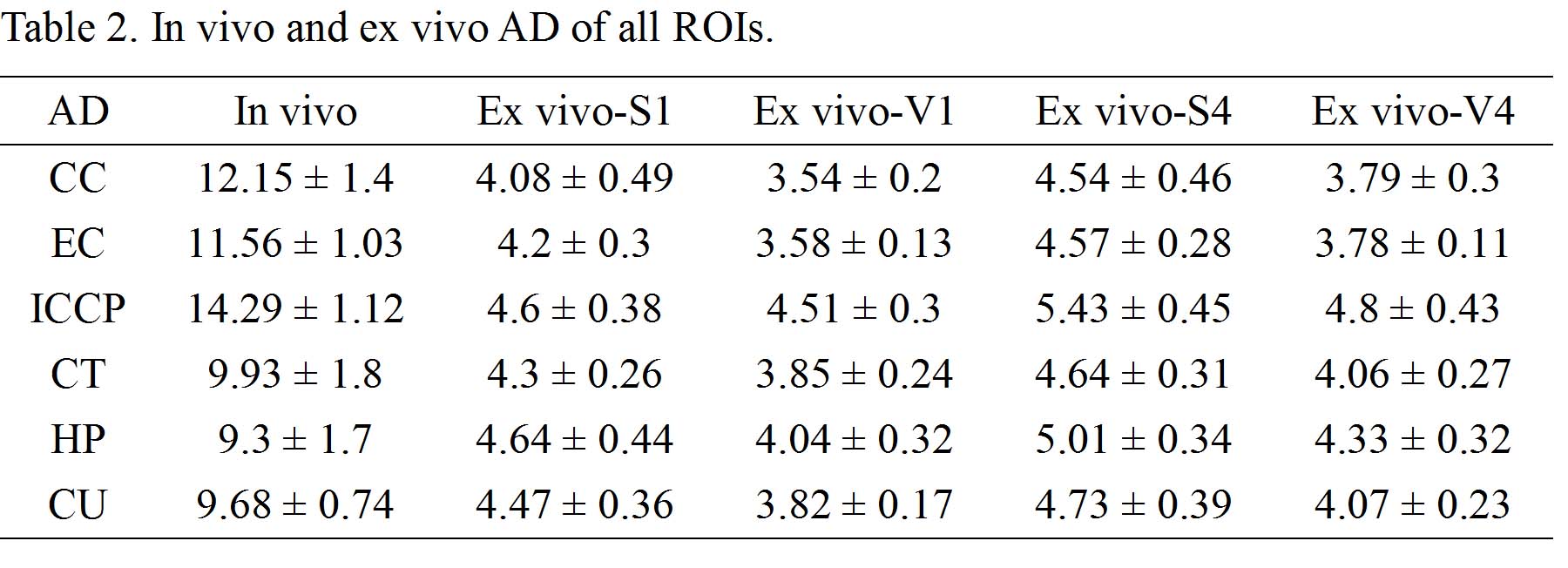
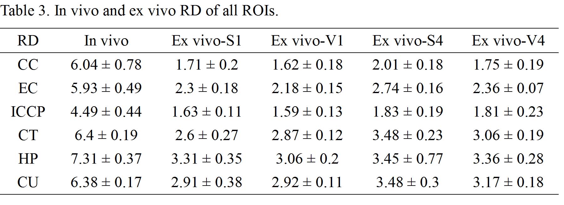
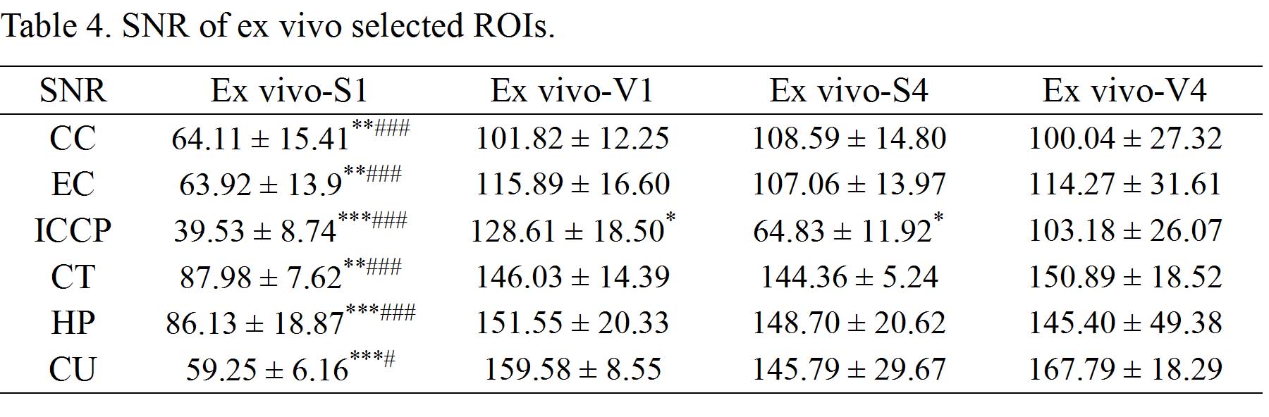
Ex vivo diffusion tensor imaging (DTI) are widely used in experimental studies for excellent images. The comparison between in vivo and ex vivo DTI has been conducted without the same scan parameters. We aimed to compare the living and fixed white and gray matters under the same condition and explore the effects of coil and signal average. Diffusivities were significant different between living and fixed brains. Coil and signal average significantly affected the signal-to-noise ratio of ex vivo white and gray matters. The results indicate that fixation and MR conditions should be considered in clinical and experimental DTI studies.

PURPOSE: To assess the effects of fixation, coil and signal average on gray and white matters using diffusion tensor imaging (DTI). METHODS: Six male SD rats underwent DTI MR for brains under conditions of in vivo with surface coil and 1 signal average (S1), ex vivo with S1 as well as volume coil with 1 and 4 averages (V1 and V4), and surface coil with 4 averages (S4). The data collected included mean diffusivity (MD), axial diffusivity (AD), radial diffusivity (RD), fractional anisotropy (FA), λ2/λ1, λ3/λ1, and λ2/λ3. Comparison of Signal-to-noise ratio (SNR) were conducted in fixed brains. Regions of interest (ROIs) included white matters (WMs) of corpus callosum (CC), external capsule (EC), and internal capsule and cerebral peduncle (ICCP), and gray matters (GMs) of cortex (CT), hippocampus (HP), and caudate putamen (CU). RESULTS: The data of diffusivities are summarized in table 1, 2, and 3. MD, AD, and RD of ex vivo ROIs with different coils and averages were lower than those of in vivo ROIs (all p < 0.001). Multi-factor ANOVA demonstrated that fixation significantly affected diffusivities of all ROIs (all p < 0.001). There were both significances and nonsignificances in comparisons of FA, λ2/λ1, λ3/λ1, and λ2/λ3 between in vivo and ex vivo from different ROIs. There were significant differences in SNR of 1 average between surface and volume coils, and in SNR of surface coil between 1 and 4 averages from all ROIs (all p < 0.05; Table 4). The multi-factor ANOVA determined that coil and average significantly affected the SNR in all the ROIs. DISCUSSION: The present study found diffusivities of brain white and gray matters reduced after fixation, which is consistent with previous studies (1-3). The principles involves structural change of proteins, tissue shrinkage, fixation processes and fixative, and temperature (4-8). Previous study confirmed the similar SNR in the cartilage with both surface and volume coils (9). However, we found higher SNR with surface coil than with volume coil, suggesting there were different coil effects on SNR in different tissues. Furthermore, there was a higher SNR from surface coil with 4 averages than with 1 average. CONCLUSION: Fixation, coil and signal average affect the DTI values, and need to take into account in clinical and experimental studies.

Acknowledgements

No acknowledgement found.

References

1. Sun SW, Neil JJ, Song SK. Relative indices of water diffusion anisotropy are equivalent in live and formalin-fixed mouse brains. Magn Reson Med. 2003;50(4):743-748.

2. Rane S, Duong TQ. Comparison of in vivo and ex vivo diffusion tensor imaging in rhesus macaques at short and long diffusion times. Open Neuroimag J. 2011;5(172-178.

3. Wu D, Xu J, McMahon MT et al. In vivo high-resolution diffusion tensor imaging of the mouse brain. Neuroimage. 2013;83(18-26.

4. Werner M, Chott A, Fabiano A et al. Effect of formalin tissue fixation and processing on immunohistochemistry. Am J Surg Pathol. 2000;24(7):1016-1019.

5. Quester R, Schroder R. The shrinkage of the human brain stem during formalin fixation and embedding in paraffin. J Neurosci Methods. 1997;75(1):81-89.

6. Wehrl HF, Bezrukov I, Wiehr S et al. Assessment of murine brain tissue shrinkage caused by different histological fixatives using magnetic resonance and computed tomography imaging. Histol Histopathol. 2015;30(5):601-613.

7. Madi S, Hasan KM, Narayana PA. Diffusion tensor imaging of in vivo and excised rat spinal cord at 7 T with an icosahedral encoding scheme. Magn Reson Med. 2005;53(1):118-125.

8. Le Bihan D. Diffusion and perfusion magnetic resonance imaging-applications to functional MRI. New York: Raven Press. 1995;

9. Bittersohl B, Huang T, Schneider E et al. High-resolution MRI of the triangular fibrocartilage complex (TFCC) at 3T: comparison of surface coil and volume coil. J Magn Reson Imaging. 2007;26(3):701-707.

Figures

All p values are less than 0.001 in the comparisons between in vivo and ex vivo MD. MD (x 10-4 mm2/s), mean diffusivity; ROI, region of interest; S1, surface coil with 1 signal average; V1, volume coil with 1 signal average; S4, surface coil with 4 signal averages; V4, volume coil with 4 signal averages; CC, corpus callosum; EC, external capsule; ICCP, internal capsule and cerebral peduncle; CT, cortex; HP, hippocampus; CU, caudate putamen.

All p values are less than 0.001 in the comparisons between in vivo and ex vivo AD. AD (x 10-4 mm2/s), axial diffusivity; ROI, region of interest; S1, surface coil with 1 signal average; V1, volume coil with 1 signal average; S4, surface coil with 4 signal averages; V4, volume coil with 4 signal averages; CC, corpus callosum; EC, external capsule; ICCP, internal capsule and cerebral peduncle; CT, cortex; HP, hippocampus; CU, caudate putamen.

There were significant differences in comparisons of in vivo and ex vivo RD (all p < 0.001). RD (x 10-4 mm2/s), radial diffusivity; ROI, region of interest; S1, surface coil with 1 signal average; V1, volume coil with 1 signal average; S4, surface coil with 4 signal averages; V4, volume coil with 4 signal averages; CC, corpus callosum; EC, external capsule; ICCP, internal capsule and cerebral peduncle; CT, cortex; HP, hippocampus; CU, caudate putamen.

Data are presented as mean ± SD. SNR, signal-to-noise ratio. ROI, region of interest; S1, surface coil with 1 signal average; V1, volume coil with 1 signal average; S4, surface coil with 4 signal averages; V4, volume coil with 4 signal averages; CC, corpus callosum; EC, external capsule; ICCP, internal capsule and cerebral peduncle; CT, cortex; HP, hippocampus; CU, caudate putamen. S1 vs. V1, V1 vs. V4, and S4 vs. V4: *, p < 0.05; **, p < 0.01; ***, p < 0.001. S1 vs. S4: #, p < 0.05; ##, p < 0.01; ###, p < 0.001.

Proc. Intl. Soc. Mag. Reson. Med. 25 (2017)
1776