1775
Detection of abnormal brain neural circuits in draxin knockout mice using DTI-MRI1Faculty of Life Sciences, Kumamoto University, Kumamoto, Japan, 2Bruker BioSpin K.K., Yokohama, Japan, 3Graduate School of Medical Sciences, Kanazawa University, Ishikawa, Japan
Synopsis
The aim of this study is to clarify the role of the axon guidance molecule Draxin in the construction of brain neural circuits by DTI-MRI. MRI was performed on draxin knockout, dra(–), and normal mice both in vivo and ex vivo. The in vivo study of dra(–) revealed that the nerve fibers in the corpus callosum did not intersect with the midline. The ex vivo study of dra(–) demonstrated that the thalamocortical nerve fibers did not extend toward the cerebral neocortex through the internal capsule. We successfully evaluated the influences caused by the Draxin loss with DTI-MRI.
Introduction
The nerve fibers in the brain are formed by an accumulation of interactions between nerve cells during the neural development process. To construct an accurate neural circuit network in the brain, the axon from one nerve cell to another target cell must extend in the correct direction. We previously identified Draxin (dorsal repulsive axon guidance protein), which is a new axon guidance molecule that shares no homology with known axon guidance molecules [1]. Draxin also reportedly acts as a nutritional factor, because draxin knockout mice show hippocampal atrophy caused by nerve cell death [2]. Therefore, Draxin is a key axon guidance molecule for the construction of brain neural circuits.Purpose
The aim of this study is to clarify the role of Draxin, by detecting the neural abnormality in draxin knockout mice with a Diffusion Tensor Imaging (DTI) method.Methods
In vivo MRI experiments were performed on both draxin knockout and normal mice under 2% isoflurane anesthesia. To obtain higher resolution, longer ex vivo MRI experiments were performed on the brains extracted from the draxin knockout and normal mice. Under anesthesia with ketamine (100 mg/kg) and xylazine (10 mg/kg), the draxin knockout and normal mice were transcardially perfused with paraformaldehyde, and after the brains were dissected from each cranium, they were stored for 24 hours at room temperature. Prior to the MRI experiments, the specimens were immersed in proton-free Fomblin Y fluid (Sigma Aldrich), in a 5 mL syringe. All MRI experiments were performed with a 7.0 Tesla BioSpec scanner and a 2-channel cryogenic probe (Bruker BioSpin). In vivo MRI data were acquired with a 2D echo planar imaging-type DTI sequence (TR 3,800 msec, TE 23 msec, matrix 128 × 128, field of view 2.2 × 2.2 cm2, resolution 172 × 172 µm2/pixel, b-value 1,582 s/mm2, 30 different gradient directions). Ex vivo MRI data were acquired with a 3D spin echo-type DTI sequence (TR 900 msec, TE 20 msec, matrix 258 × 134 × 175, field of view 1.44 × 7.50 × 9.80 cm3, resolution 56 × 56 × 56 µm3/pixel, b-value 1,000 s/mm2, 30 different gradient directions). Fractional anisotropy (FA) analyses and tractography were performed with the ParaVision software (Bruker BioSpin) and the Diffusion Toolkit/TrackVis software [3], respectively.Results and Discussion
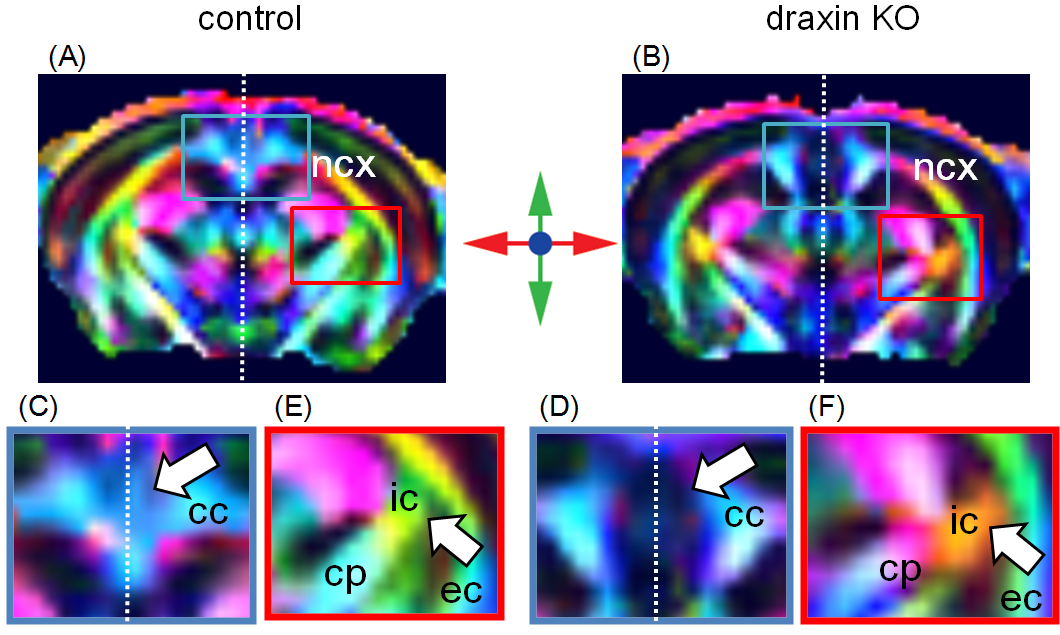
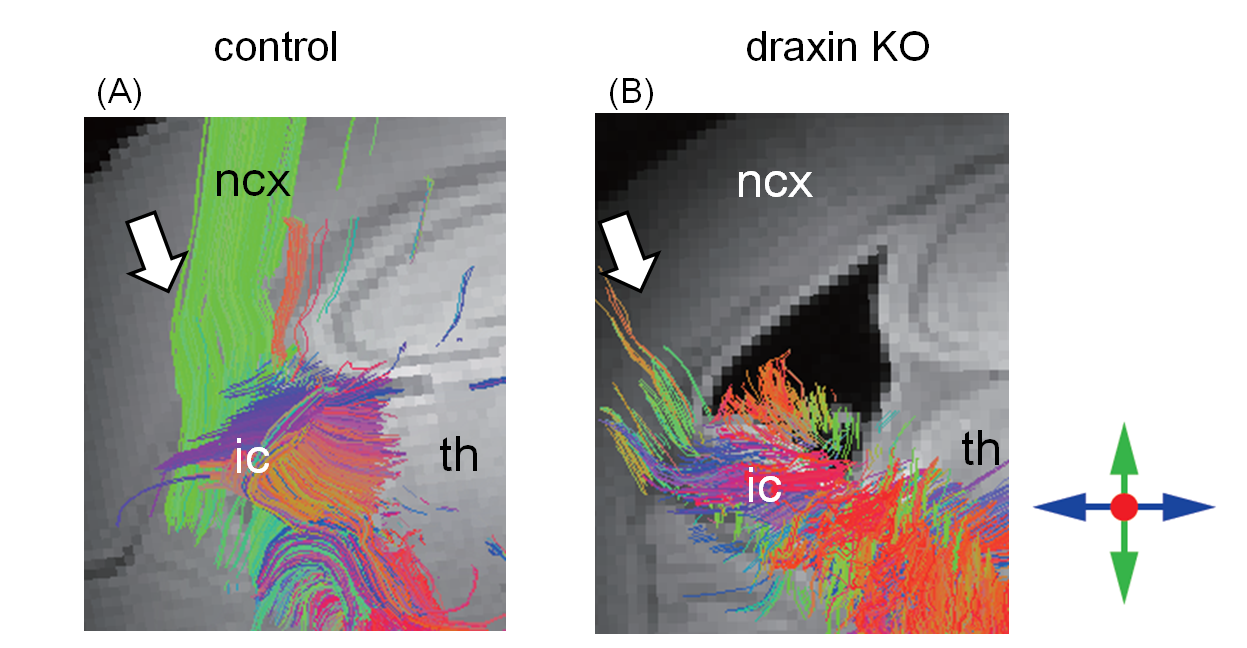
FA analyses were applied to the in vivo data of the draxin knockout and normal mice (Fig. 1). The normal mouse data showed that the nerve fibers in the corpus callosum intersect with the midline (Fig. 1A and C), while the draxin knockout mouse data revealed an abnormality, in that the fibers did not intersect with the midline (Fig. 1B and D). The normal mouse data also showed that the nerve fibers in the internal capsule extended in the top–bottom direction (Fig. 1E), while the draxin knockout mouse data showed the fibers extended in the right–left direction (Fig. 1F). To obtain 3D information with higher resolution, tractography was performed in the extracted brains of the draxin knockout and normal mice (Fig. 2). The normal mouse brain data showed that the thalamocortical nerve fibers extended toward the cerebral neocortex through the internal capsule (Fig. 2A), while the draxin knockout mouse brain data showed that the fibers did not extend toward the cerebral neocortex (Fig. 2B). These results corresponded with the results of our previous haematoxylin and eosin-staining experiments [4]. The shrinked hippocampus was also confirmed in the ex vivo anatomical images of draxin knockout mice, in comparison with the images of normal mice (Fig. 2). It is noteworthy that much more 3D information about the brain neural fiber track in the draxin knockout mice was obtained in this study.Conclusions
We successfully detected a neural abnormality in the draxin knockout mice, both in vivo and ex vivo.Perspective
In the future, we plan to clarify the abnormal development of the brains of young to adult draxin knockout mice by DTI-MRI.Acknowledgements
No acknowledgement found.References
[1] Islam, S. M. et al., Science 323, 388–393 (2009)
[2] Zhang, S. et al., Neurosci. Res. 66, 53–61 (2010)
[3] Wang, R. et al., Proc. Intl. Mag. Reson. Med. 15, 3720 (2007)
[4] Shinmyo, Y. et al., Nature Commun. 6, 10232 (2015)
Figures