1766
Diffusion Imaging Reveals White Matter Damage in Ice Hockey Players for Up To Two Months Post-Concussion1University of British Columbia, Vancouver, BC, Canada, 2Pediatrics, University of British Columbia, Vancouver, BC, Canada
Synopsis
Traumatic brain injury (TBI) is a leading cause of disability in adults. More sophisticated methods are required in order to understand its underlying pathophysiology and disease progression. Recent interest in an assumption-free DTI analysis, diffusion entropy (DE), has produced some promising results for investigating white matter (WM) and grey matter (GM) microstructure. We set out to test DE against the much more common fractional anisotropy (FA) analysis, in both WM and GM, in 45 hockey players over one season. 11 players sustained a concussion and were scanned at 72 hours, 2 weeks, and 2 months post-injury.
Introduction
Traumatic brain injury (TBI) is a leading cause of disability in adults, making up 9% of all trauma hospital admissions in Canada [1]. While the mild form of TBI – concussions – have historically been treated as non-critical, more recent developments have lead to an understanding of the very serious short and long term negative health effects they can have [2-5]. As the diagnosis and management of the injuries are largely based on self-reported symptoms, a more sophisticated method is required in order to understand the underlying pathophysiology and disease progression. Recent interest in an assumption-free DTI analysis, diffusion entropy (DE), has produced some promising results for investigating white matter (WM) and grey matter (GM) microstructure [6,7,8]. We set out to test DE against the much more common fractional anisotropy (FA) analysis, in both WM and GM, with respect to probing mTBI damage in university varsity hockey players over one season of athletic play.Methods
Subjects: This study used data acquired from an earlier completed study, and is thus retrospective. Twenty five male and twenty female amateur ice hockey players (mean age ± SD) = 21.2±3.1years) from two Canadian Interuniversity Sports teams participated in the study. All players were scanned (baseline) before the beginning of the hockey season (September). An independent physician was on hand for all games, in order to diagnose concussions. If a player was concussed, they were then scanned at 72hrs, 2weeks, and 2months after their concussion.
MRI: MRI data were acquired on a Philips Achieva 3T scanner. All subjects underwent the following set of scans: sagittal 3D T1-weighted scan (TR/TE/flip angle=8.1ms/3.7ms/6°; acquisition matrix/FOV/acquired voxel size/reconstructed=256x256x160/256x256x160mm$$^3$$/1x1x1mm3/1x1x1mm3); and DTI scan (TR/TE/flip angle=7015ms/60ms/90°; acquisition matrix/FOV/acquired voxel size/reconstructed/slice thickness=100x99/224x224mm2/2.2x2.2mm2/2x2mm2/2.2mm; b0=0, b1=700sec/mm2, 60 non-collinear directions).
Data Processing: DTI data was eddy current corrected, brain extracted, and FA values calculated using FSL. DE values were calculated from the eddy current corrected diffusion data based on Equation (1) using in-house written software (MATLAB). Briefly, for each subject, and then for each voxel, signal attenuation values for every direction in the voxel were binned into 60 bins between 0 and 1. Probability density functions for each bin were calculated. Substituting these values into Equation (1) gave the DE value.
Equation(1): $$ DE(x_i)=-\sum_{x_{i} \epsilon k} {(p(x_i)log(p(x_i))}$$
Statistics: JHU atlas masks of the genu and splenium were projected onto FA and DE volumes, and averaged for each subject. Subcortical grey matter regions (thalamus and hippocampus) were segmentedfrom the 3D T1 images. Linear mixed effects models (R: lme450) were used to analyze the relationship between FA or DE and time. Time was used as a fixed effect, with intercepts for subjects as random effects in the model (e.g.: m <- lmer(de ~ time + (1|subject), data=icehockey). Post-hoc analysis was performed in order to determine which time-points were significantly different from baseline (pair-wise t-tests, p<0.05, corrected for multiple comparisons using the Holm-Bonferonni method). This analysis was performed for the four ROIs (gCC, sCC, thalamus, and hippocampus).
Results
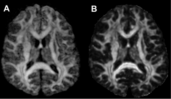
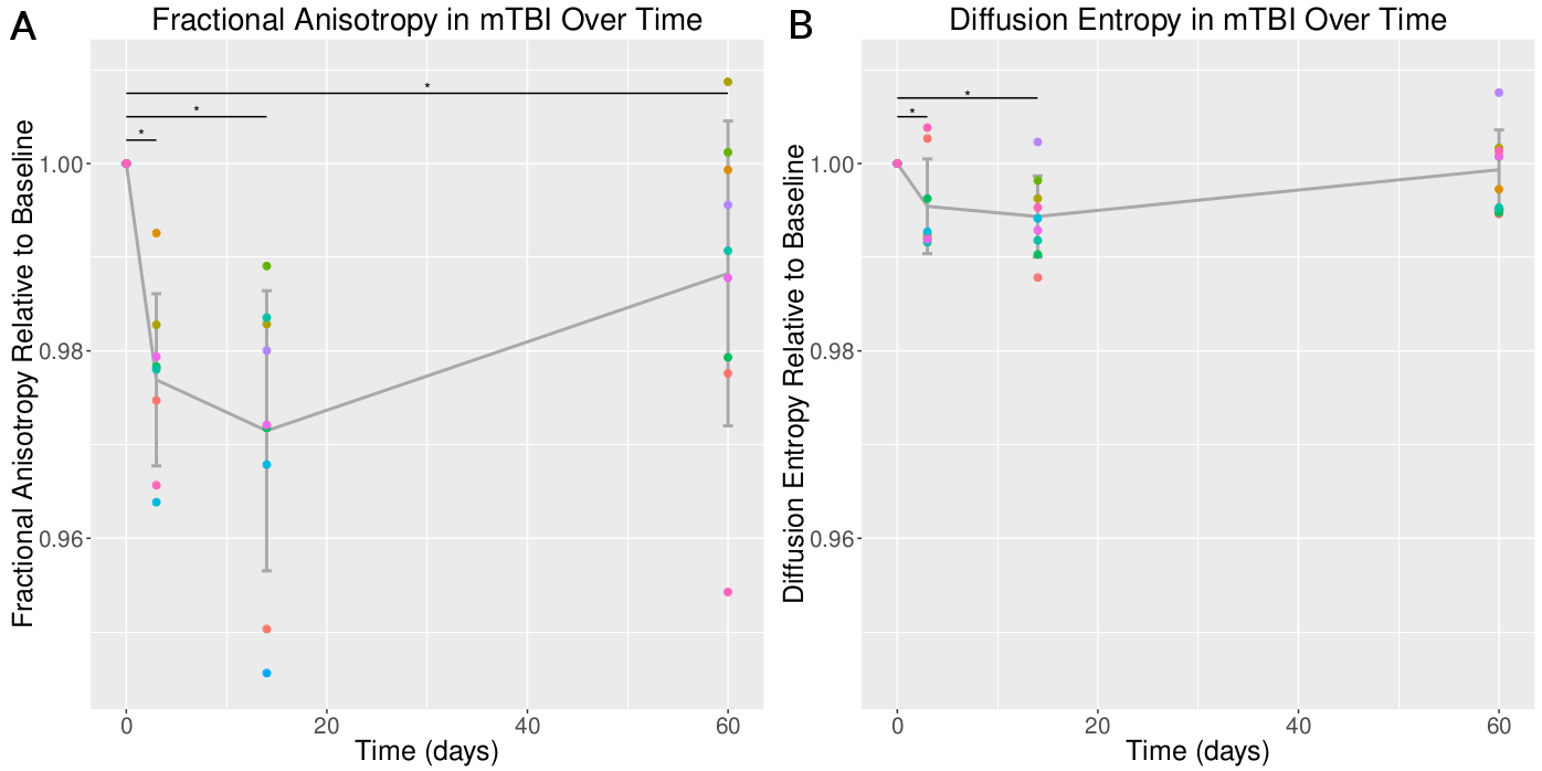
11 players sustained a concussion during competition, and were scanned at 72 hours, 2 weeks, and 2 months post-injury. DE and FA maps differed most with respect to GM contrast (Fig1). Linear mixed effects models revealed significant longitudinal differences in FA and DE values after concussion in the gCC, but not in the sCC, thalamus or hippocampus. In the gCC, post-hoc analysis revealed differences after 3, 14 and 60 days compared to baseline post concussion in FA values, while differences were only found after 3 and 14 days in DE values (Fig2).Discussion
We report the first large-scale comparison of DE and FA values in measuring micro-structural changes post-concussion in human subjects. Unlike TBI studies performed in rats, we did not find a benefit in using DE over FA, in both white matter and grey matter. Our results show that both DE and FA exhibited decreased values in gCC after sustaining a concussion, at 3 days and at 2 weeks. FA, however, still exhibited decreased values after 60 days. Our findings are not conclusive, however, and further studies should be performed, as there are some hints that DE be beneficial (cortical GM, and WM recovery). With respect to how these findings could effect return-to-play in sport-related concussions, our analysis suggests that, despite recovering in terms of executive and cognitive symptoms, WM may remain damaged, and in a vulnerable period of recovery. This period extended to 2 months, and possibly longer (as we did not collect data past this time-point), suggesting that the window of elevated vulnerability to repeated impacts extends well beyond functional recovery.Acknowledgements
Author contributions: A.R., J.T. and D.L. designed the study. A.R. and D.L. designed the imaging protocol. S.D. collected data and helped coordinate the study. M.J., and A.M.W. performed data analysis under the supervision of A.R. A.M.W. wrote the manuscript. E.H.-T. edited and provided critical input to the manuscript. All authors interpreted the data. All authors had full access to the data, and helped critically revise the manuscript before reviewing and approving the final version. Competing interests: The authors declare that they have no competing interests. Data and materials availability: Data is available and may be provided under the transfer policies of UBC.[
References
[1] Canadian Institute for Health Information (CIHI). Head Injuries in Canada: A Decade of Change (1994-1995 to 2003-2004) [Internet]. 2006 [cited 2016 Apr 8]. Available from: https://secure.cihi.ca/estore/productFamily.htm?pf=PFC1360&lang=en&media=0
[2] Guskiewicz KM, McCrea M, Marshall SW, et al. Cumulative effects associated with recurrent concussion in collegiate football players: The ncaa concussion study. JAMA. 2003 Nov 19;290(19):2549–55.
[3]
Centers for Disease Control and Prevention (CDC), National Center for
Injury Prevention and Control. Report to Congress on mild traumatic
brain injury in the United States: steps to prevent a serious public
health problem. Centers for Disease Control and Prevention; 2003.
[4]
Belanger HG, Curtiss G, Demery JA, Lebowitz BK, Vanderploeg RD.
Factors moderating neuropsychological outcomes following mild
traumatic brain injury: a meta-analysis. J Int Neuropsychol Soc JINS.
2005 May;11(3):215–27.
[5]
Ryan LM, Warden DL. Post concussion syndrome. Int Rev Psychiatry
Abingdon Engl. 2003 Nov;15(4):310–6.
[6]
Fozouni N, Chopp M, Nejad-Davarani SP, Zhang ZG, Lehman NL, Gu S, et
al. Characterizing brain structures and remodeling after TBI based on
information content, diffusion entropy. PloS One. 2013;8(10):e76343.
[7]
Metwalli NS, LaConte SM, Hu XP. An information theoretic approach
characterizing diffusion anisotropy in diffusion-weighted magnetic
resonance images. Conf Proc Annu Int Conf IEEE Eng Med Biol Soc IEEE
Eng Med Biol Soc Annu Conf. 2006;1:2260–3.
[8]
Li L, Chopp M, Ding G, Qu C, Nejad-Davarani SP, Davoodi-Bojd E, et
al. Diffusion-Derived Magnetic Resonance Imaging Measures of
Longitudinal Microstructural Remodeling Induced by Marrow Stromal
Cell Therapy after Traumatic Brain Injury. J Neurotrauma. 2016 May 13.
Figures