1736
A Fractional Motion Diffusion Model for a Twice-Refocused Spin Echo Pulse Sequence: Analytical Expression and in vivo Demonstration1Center for MR Research, University of Illinois at Chicago, Chicago, IL, United States, 2Departments of Radiology, Neurosurgery, and Bioengineering, University of Illinois at Chicago, Chicago, IL, United States
Synopsis
The anomalous diffusion behavior of biological tissue has been investigated by several studies using non-Gaussian diffusion models. Among these, the fractional motion (FM) model has attracted an intense interest by the biophysics community and was recently been introduced to the MR community. The current diffusion signal formulism based on the FM model is limited to a simple Stejskal-Tanner gradient despite the widespread use of the eddy-current-resistant gradient waveform with the twice-refocused spin echo (TRSE) technique. In this study, we theoretically derive a formulism based on the FM diffusion model to characterize anomalous diffusion using a TRSE sequence, and experimentally validate it on healthy human brain in vivo.
Introduction
It has been well recognized that the diffusion process at high b-values does not follow the classical Gaussian distribution in biological tissues due to structural complexity, heterogeneity, and other factors. Several studies have investigated this “anomalous” behavior through theoretical models that aim to reveal the actual physical makeup of the underlying tissue structure and complexity in relation to diffusion “abnormalities”. Among several anomalous diffusion models, the continuous-time random walk model (CTRW)1,2 (together with its associated fractional order calculus model3-5) and the fractional motion (FM) model6,7 have been actively pursued by the biophysics community to understand the intrinsic diffusion process in biological systems. While the CTRW model has already been applied to MR diffusion imaging for several years1,2, the FM model was introduced to the MR community only very recently6,7. The existing FM model is limited to a simple Stejskal-Tanner gradient. On human MR scanners, however, an eddy-current-resistant gradient waveform using twice-refocused spin echo (TRSE)8 is often used, yet no analytical expression exists to describe the anomalous diffusion MR signal under the FM model. The purpose of this study is to develop a formulism for TRSE-specific FM model and demonstrate its applicability to human brain in vivo.Theory
The FM diffusion model is based on Langevin motion, which is a unified statistical form for motions with power-law Fourier and kernel functions9. Considering correlated consecutive diffusion increments as a result of power law, the signal attenuation due to diffusion takes the following form: $$S/S_0=exp(-D_{φ,ψ} \int_{0}^{T_0}|F(t)|^φ dt), [1] $$ $$F(t)=\int_{t}^{T_0} γG(τ) (τ-t)^{H-1/φ}dτ, [2]$$ where Dφ,ψ is an anomalous diffusion coefficient (mm2/s), and φ and ψ are the parameters governing the variance and correlation properties of the increments of diffusion. The term, $$$(τ-t)^{H-1/φ}$$$, is a power law memory kernel7,9 with the Hurst exponent $$$H=ψ/φ$$$10. For the TRSE implementation that balances the concomitant gradient fields11, the gradient waveform (Fig. 1) consists of two bipolar gradients of length $$$δ_1$$$ and $$$δ_2$$$and, an inter-gradient separation, s. The diffusion-weighted (DW) signal attenuation for a TRSE sequence can be computed by obtaining F(t) in Eq.[2] for the gradient waveform, leading to the following signal equation: $$S/S_0=exp(-η^{''}D_{φ,ψ}(γG)^{φ}Δ_1^{φ+ψ}), [3]$$ where $$$Δ_1=δ_1+s$$$. The analytical expression of a dimensionless factor, $$$η^{''}$$$, is shown in Fig.2.
Methods
Image acquisition: DW imaging experiments were performed on a 3T GE MR750 scanner. A TRSE sequence shown in Fig. 1 was used to acquire DW images from six healthy human brains. A total of 15 b-values (01, 101, 201, 501, 1001, 2002, 4002, 7004, 10004, 15008, 20008, 250016, 300016, 350016, 400016 s/mm2, with the subscripts denoting the NEXs, was used. Trace-weighted images were obtained to minimize the effect of diffusion anisotropy. The other sequence parameters were δ1=10.8ms, δ2=23.1ms, s=6.9ms, TR/TE=4200/106ms, reconstructed matrix size=256×256, slice thickness=3mm, scan time=24 minutes.
Analysis: The multi-b-value DW images were fitted to Eq.[3] to estimate the FM parameters, Dφ,ψ, φ, ψ, by using a non-linear least squares algorithm. The FM parameters were evaluated in specific regions of interest (ROIs) in gray matter (GM), white matter (WM), and cerebrospinal fluid (CSF) as shown in Fig.3a.
Results
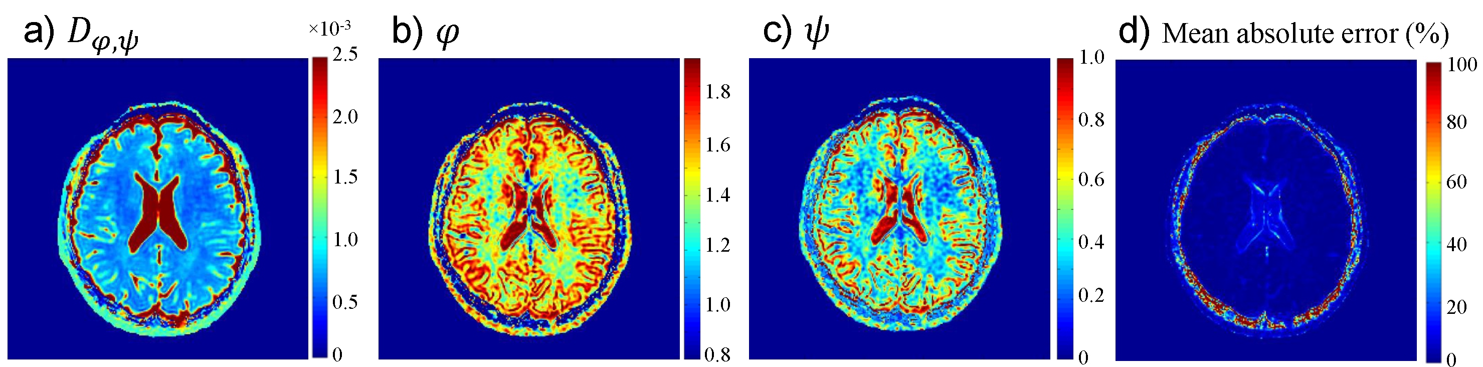
Figure 3b illustrates the agreement between the signal attenuation and the fitting curves using the FM model on the ROIs given in Fig.3a. Figure 4 summarizes the sample means ($$$\overline{x}$$$) and standard deviations (σ) of the FM parameters,Dφ,ψ, φ, ψ, as well as the mean absolute error (MAE) of the fitting in the three representative ROIs. The results showed that the FM parameters exhibited good contrast for the different tissues (i.e., different $$$\overline{x}$$$) as well as low variation within a tissue (i.e., σ). In addition, the MAEs ( $$$\overline{x}$$$ and σ) were small in all regions. These good performances were further illustrated in Fig. 5 where spatially resolved maps of Dφ,ψ, φ, ψ, and MAE obtained from voxel-by-voxel fitting of Eq.[3] for a representative subject is displayed. Both Figs. 4 and 5 illustrate that the FM model well described the diffusion signal obtained from a TRSE sequence.
Discussion and Conclusion
We have theoretically derived a new expression for the FM diffusion model to characterize the DW signal in a TRSE pulse sequence. Our experimental results showed that this theoretical formulism can accurately describe the high-b-value diffusion behavior in healthy brain tissues at b-values. As high b-value diffusion imaging is susceptible to increased eddy currents perturbations, the TRSE pulse sequence is particularly important to provide immunity to eddy currents for anomalous diffusion studies. As such, the formulism reported herein is expected to enable a more extensive exploration of the new FM diffusion model for anomalous diffusion imaging.Acknowledgements
This work was supported in part by NIH 1S10RR028898. We thank Drs. Kejia Cai, Frederick C. Damen, and Richard L. Magin for valuable discussions.References
[1] Ingo C, Magin RL, Colon-Perez L, et al. On random walks and entropy in diffusion-weighted magnetic resonance imaging studies of neural tissue. Magn Reson Med. 2014;71(2):617–27.
[2] Karaman MM, Sui Y, Wang H, et al. Differentiating low- and high-grade pediatric brain tumors using a continuous-time random-walk diffusion model at high b-values. Magn Reson Med. 2015; 76:1149-1157.
[3] Magin RL, Abdullah O, Baleanu D, et al. Anomalous diffusion expressed through fractional order differential operators in the Bloch-Torrey equation. J Magn Reson Im. 2008;190:255–270.
[4] Zhou XJ, Gao Q, Abdullah O, Magin RL. Studies of anomalous diffusion in the human brain using fractional order calculus. Magn Reson Med. 2010;63:562–569.
[5] Sui Y, He W, Damen FW, Wanamaker C, Li Y, Zhou XJ. Differentiation of Low- and High- Grade Pediatric Brain Tumors with High b-Value Diffusion weighted MR Imaging and a Fractional Order Calculus Model. Radiology. 2015;277(2):489–496.
[6] Karaman MM, Wang H, Sui Y, et al. Fractional Motion Diffusion Model for Grading Pediatric Brain Tumors. Neuroimage Clin. 2016: 12:707-714.
[7] Fan Y, Gao JH. Fractional motion model for characterization of anomalous diffusion from NMR signals. Phys Rev E. 2015;92(1):012707.
[8] Reese TG, Heid O, Weisskoff RM, et al. Reduction of eddy-current-induced distortion in diffusion MRI using a twice-refocused spin echo. Magn Reson Med 2003;49:177–182.
[9] Eliazar II, Shlesinger MF. Fractional motions. Phys Rep. 2013;527(2):101–29.
[10] Hurst HE. Long-term storage capacity of reservoirs. Trans Am Soc Civ Eng. 1951;116:770-799.
[11] Gao Q, Srinivasan G, Magin RL, Zhou XJ. Anomalous diffusion measured by a twice-refocused spin echo pulse sequence: Analysis using fractional order calculus. J Magn Reson Imaging. 2011;33(5):1177-1183.
Figures