1732
Diffusion-weighted MR imaging of kidney after administration of 2 types of iodinated contrast medium: a time course study in CIN animal model1Department of Radiology, Peking University First Hospital, Beijing, People's Republic of China
Synopsis
DWI is a preeminent noninvasive method to quantify renal function, which may be helpful to understand the pathogenesis of CIN. Our time course study indicates that the iodinated contrast medium can induce some affect to the different zone of kidney. And some differences do exist on the renal transport function after the two kinds of iodinated CM administration.
Introduction and Purpose
Usage of iodinated contrast medium (CM) during radiological procedure may cause contrast induced nephropathy (CIN), which has drawn an increasing attention in the whole world. The purpose of this study is to find out the chronological effect of 2 types of iodinated contrast medium in intrarenal water diffusion in CIN animal model by diffusion-weighted MR imaging.Method
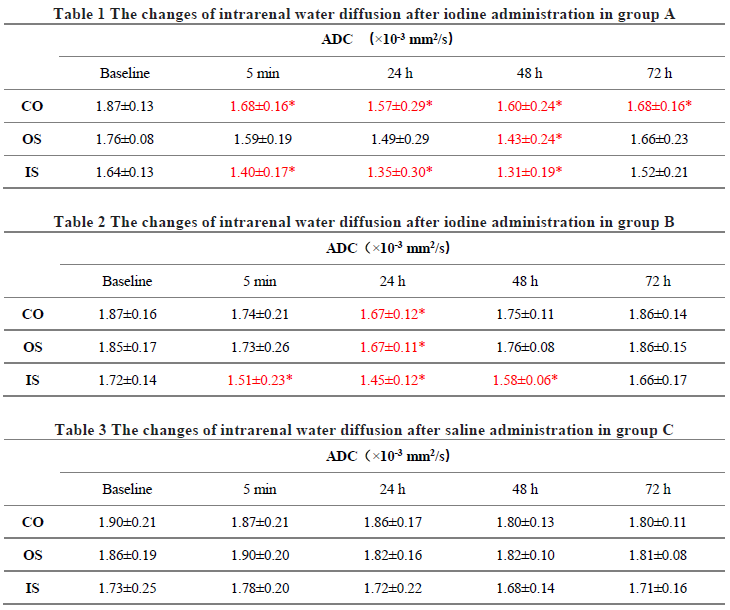
Seventeen New Zealand white rabbits were divided into 3 groups: Group A (Iodixanol-270, n=6), Group B (Iohexol-350, n=6), Group C (0.9% saline, n=5). Respiratory anesthesia was used. MR studies were conducted in a 3.0T GE MR scanner with a knee coil. A sequential DWI (0 and 800 s/mm2) was performed to estimate the intrarenal apparent diffusion coefficient (ADC) at 24h before and 5min, 24h, 48h, 72h after contrast or saline administration. Iodinated contrast agent with a dose of 1380 mg I/kg were injected to the experimental groups (Group A and B), and 0.9% saline with a dose of 5.1 ml/kg (to keep the liquid load equal) was administrated to the control group (Group C). ADC values obtained in the cortex (CO), outer stripe of the outer medulla (OS), inner stripe of the outer medulla (IS) were grouped into 5 time-points : “baseline”, “5min”, “24h”, “48h”, “72h” respectively. Then at last the pathology was obtained. Paired sample t-test was performed to test the change of intrarenal water diffusion after iodine administration within each group, and One-Way ANOVA was performed to test the response among the 3 groups in each time-point.Results
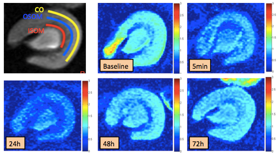
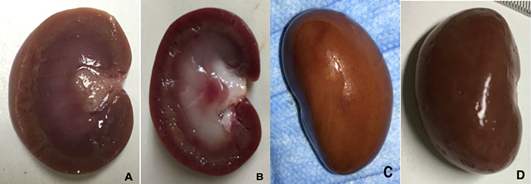
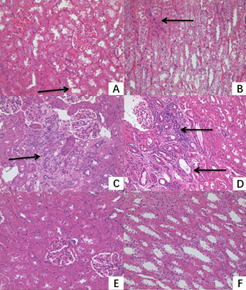
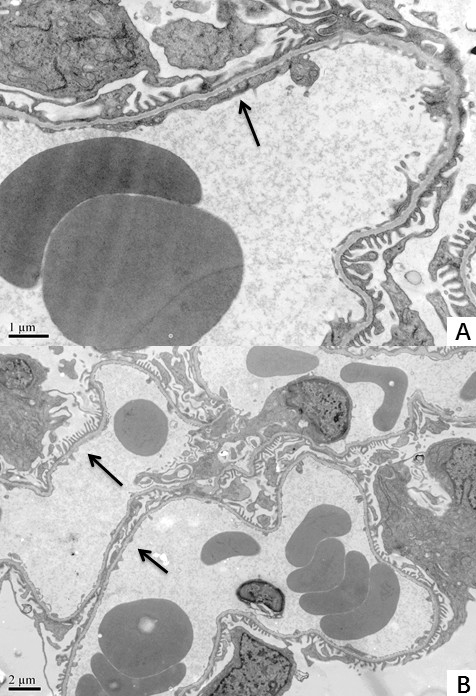
The iopamidol induced more serious decrease in ADCs in the CO, OS and IS of the kidney compared with the saline, which was shown in Table 1, 2 and 3. Among the 3 groups, there was no significant difference in the CO, OS and IS before administration. But Iodixanol-270 produced more damage in the CO while both Iodixanol-270 and Iohexol-350 induced more damage in the IS 5min, 24h, 48h after administration compared with Group C (Figure 1). As for pathology, significant differences were detected after large dose iopamidol administration in the iodine group (Figure 2, 3, 4). Iopamidol produced moderate to severe tubular epithelial cellular swelling, vacuolar degeneration, necrosis and shedding into the tubular lumens. Focal or multifocal lymphocytes and mononuclear cells infiltration in the interstitium appeared in most of the kidneys. The pathologic changes in Group B was more serious than that in Group A. For the kidneys in the saline group (Group C), light microscopic examinations showed that there was no obvious pathological change in the glomeruli.Discussion
In this study, rabbits with CIN are animal models. Usage of iodinated contrast medium during radiological procedure may cause CIN. In human, CIN is characterized by an increase in the serum creatinine (Scr) level of at least 0.5 mg/dl or 25% compared to baseline values in the absence of an alternative etiology1. CIN is more and more significant with the increasing use of enhanced CT and interventional diagnosis and treatment. DWI is a preeminent noninvasive method to quantify renal function, which may be helpful to understand the pathogenesis of CIN. Our time course study indicates that the iodinated contrast medium can induce some affect to the different zone of kidneys. While some differences do exist on the renal transport function in the two kinds of iodinated CM by DWI. As we know, the osmotic pressure of Iodixanol-270 (290 mOsm/kgH2O) is much lower than that of Iohexol-350 (844 mOsm/kgH2O). At the same time, the viscosity of Iodixanol-270 (11.3 mPa·s) is also lower than that of Iohexol-350 (11.6 mPa·s). Which factor is the most important? Osmotic pressure? Viscosity? Or other factors? More research is needed to figure out which kine of contrast medium is less harmful to human.
Conclusion
The time course study indicates that some differences do exist on the renal transport function after the two kinds of iodinated CM administration, which can be detected by DWI scan and be confirmed by pathology.Acknowledgements
No acknowledgement found.References
1. Thomsen HS. Guidelines for contrast media from the European Society of Urogenital Radiology. AJR American journal of roentgenology. 2003;181(6):1463-1471.Figures