1725
Thalamocortical neural responses to whole body heat exposure: A resting-State Functional MRI study1Jinan Military General Hospital, Jinan, People's Republic of China, 2GE Healthcare China
Synopsis
In this study, we replicated previous findings about spatially distinct thalamic correlations with cortical regions of interest. Using bi-directional functional analysis, we firstly clarified cortical-subcortical activity during hyperthermic condition by thalamocortical functional connectivity. Specifically, we found weakening connectivity of cortical fronto-polar/anterior cingulate cortex and prefrontal areas with the corresponding thalamic nuclei. On the contrary, the motor/premotor and somatosensory cortical subdivisions showed increased connectivity with thalamic nuclei. The thalamic pulvinar showed reduced connectivity with bilateral superior and middle temporal pole, but increased connectivity with bilateral middle and inferior temporal gyrus.
Purpose
Passive hyperthermia is a potential risk factor to human in many extreme work environments, such as a product manufacturing plant, coal mine, military operation, firefighting and outdoor sports[1; 2]. Previous studies have shed light on the significant effects of passive hyperthermia on human cortical-cortical activity during resting-state, visual attention network task, working memory task[3-5]. However, the potential principles of human cognition and behavior deteriorations were still not clearly known. These previous studies focused on cortical activity related to high level cognition and behavior, but the relationships with subcortical activity which was mainly related to low level thermal sensation and regulation were neglected.With unique cytoarchitecuture and firing patterns in human brain, the thalamus acts as a core structure which contains wide spread connections with distinct zones of the cerebral cortex, named thalamocortical network previously [6; 7]. The thalamus acts as a key hub of spinothalamic tract and thalamocortical radiations, involving with both sensory information transmission from bottom neural activity and cognition processing from top neural activity[8].With the development of functional imaging and fiber tracts, the thalamocortical connectivity provide a valuable approach for imaging cortical-subcortical neural activity[9]. Therefore, the aim was to examine the pattern of thalamocortical connectivity would be disrupted during whole body heat exposure for an hour.Methods
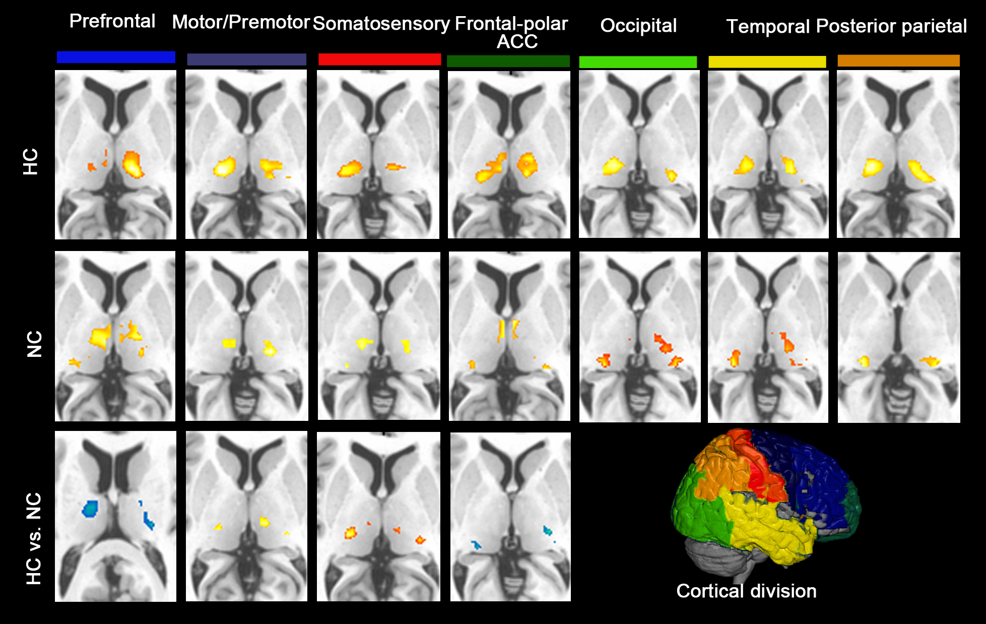
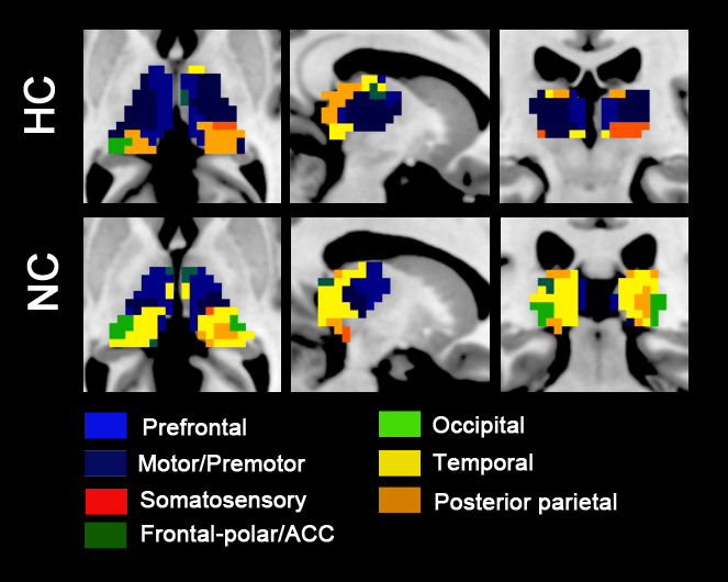
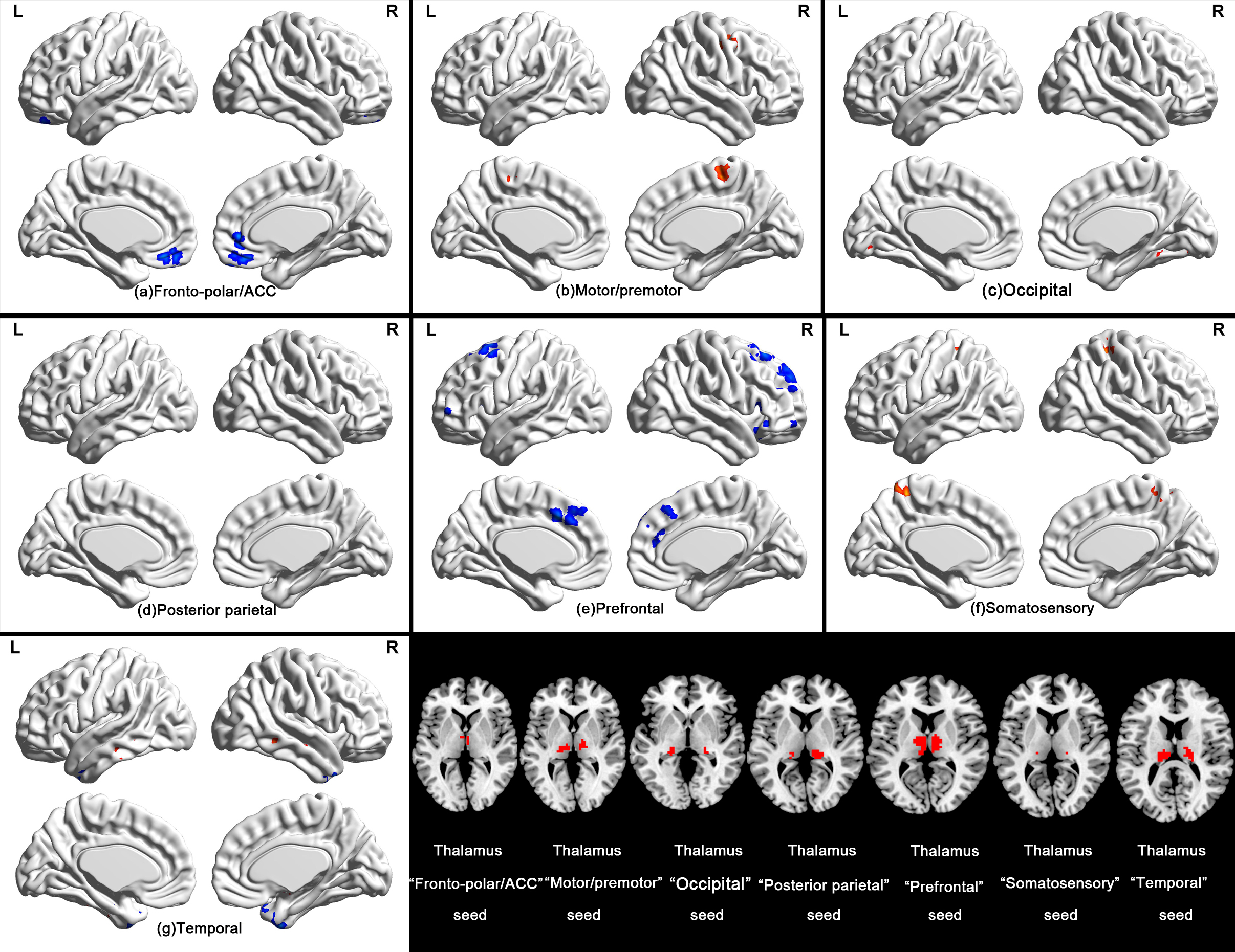
Two thermal conditions were performed on 18 participants, a normal control (NC) condition (50 min, 25 ◦C) and a hyperthermia condition(HC) (50 min, 50 ◦C), in an environmental chamber in a counterbalanced order with 3~7 days apart. After that, they were asked to move into the scanner room. During MRI scanning, heat procedure was continued. The cortex was divided into seven cortical regions based on anatomical landmarks, and the thalamus was parceled into functional ROIs using the winner take all strategy[9]. We performed thalamocortical connectivity analysis in the wise of bidirectional ROI-voxel manner. Both of the cortico-thalamus and thalamic-cortical connections were calculated.Results
During the cortico-thalamic correlation analysis, we identified significantly altered correlations of cortical ROIs (prefrontal, motor/premotor, somatosensory, frontal-polar/anterior cingulate cortex) with the thalamus (Figure 1). The prefrontal subdivision was robustly connected to medial dorsal nucleus, but decreased connectivity was found during hyperthermic condition. Increased thalamic connectivity was showed with somatosensory and motor/premotor seeds in ventral lateral nucleus during hyperthermic condition. The frontal-polar and ACC showed decreased connectivity with pulvinar was found during hyperthermic condition. Using the winner take all approach, we found the altered spatial extent and intensity of cortical ROIs (except occipital) to thalamus (Figure 2). During the thalamic-cortical correlation analysis, thalamus “fronto-polar/ACC” seed connectivity with orbital medial superior frontal gyrus and ACC was reduced during hyperthermic condition. Furthermore, participants exhibited reduced thalamus “prefrontal” seed connectivity with bilateral superior/middle frontal gyrus during hyperthermic condition. While thalamus “motor/premotor” and “somatosensory” seeds showed hyperconnectivity with with bilateral precentral gyrus, paracentral lobule, right middle frontal gyrus and postcentral gyrus (Figure 3).Conclusions
These thalamocortical connectivity alterations might reflect hyperthermia-induced thermal sensation, regulation and neural behavior changes underlying cortical-subcortical activity. These distinct thalamocortical connectivity changes revealed that the different thalmocortical pathways exist for hyperthermia-induced physiological and psychological disruptions.Acknowledgements
No acknowledgement found.References
1. Hocking C, Silberstein RB, Lau WM, et al. Evaluation of cognitive performance in the heat by functional brain imaging and psychometric testing [J]. Comp Biochem Physiol A Mol Integr Physiol, 2001, 128(4):719-734
2. Hancock PA, Vasmatzidis I. Effects of heat stress on cognitive performance: the current state of knowledge [J]. Int J Hyperthermia, 2003, 19(3):355-372
3. Sun G, Qian S, Jiang Q, et al. Hyperthermia-induced disruption of functional connectivity in the human brain network [J]. PLoS One, 2013, 8(4):e61157
4. Liu K, Sun G, Li B, et al. The impact of passive hyperthermia on human attention networks: An fMRI study [J]. Behav Brain Res, 2013, 243C220-230
5. Jiang Q, Yang X, Liu K, et al. Hyperthermia impaired human visual short-term memory: an fMRI study [J]. Int J Hyperthermia, 2013, 29(3):219-224
6. Zhang D, Snyder AZ, Shimony JS, et al. Noninvasive functional and structural connectivity mapping of the human thalamocortical system [J]. Cereb Cortex, 2010, 20(5):1187-1194
7. Woodward ND, Karbasforoushan H, Heckers S. Thalamocortical dysconnectivity in schizophrenia [J]. Am J Psychiatry, 2012, 169(10):1092-1099
8. Yuan R, Di X, Taylor PA, et al. Functional topography of the thalamocortical system in human [J]. Brain Struct Funct, 2015. 10.1007/s00429-015-1018-7
9. Woodward ND, Heckers S. Mapping Thalamocortical Functional Connectivity in Chronic and Early Stages of Psychotic Disorders [J]. Biol Psychiatry, 2016, 79(12):1016-1025
Figures