1712
Increased inter-hemispheric functional connectivity in restless legs syndrome using voxel-mirrored homotopic connectivity1MR Research China, GE Healthcare, Shanghai, People's Republic of China, 2Radiology, Shanghai First People's Hospital, Shanghai, People's Republic of China, 3Neurology, Shanghai First People's Hospital, Shanghai, 4MR Research China, GE Healthcare, Beijing, People's Republic of China
Synopsis
This preliminary study used voxel- mirrored homotopic connectivity (VMHC), a novel resting-state fMRI parameter to investigate inter-hemispheric functional activity changes in restless legs syndrome (RLS). Ten RLS patients and ten age- and gender-matched healthy controls were recruited for comparison. The RLS group showed increased VMHC in the amygdala, putamen and insula, as compared to normal controls. Increased VMHC in the insular cortex and putamen might reflect their functions in perception and motor control. Increased VMHC in the amygdalae could be relevant to the disturbed sleep at night, considering its primary role in the processing of memory and emotion.
Purpose
Restless legs syndrome (RLS), a common neurological disorder, is presumed to be caused by iron deficiency and dopaminergic dysfunction within the brain. The patients suffer an urge to move the legs and paraesthesia deep in the legs, which could cause severe sleep disturbances at night.1 The previous resting-state fMRI studies often used local parameters, for example, Amplitude of Low Frequency Fluctuation (ALFF) and Regional homogeneity (ReHo) to investigate local spontaneous brain activities, which might be incapable to reflect the changes of the whole brain functional activity.2 Voxel-mirrored homotopic connectivity (VMHC), a novel resting-state fMRI parameter has been developed to calculate the functional connectivity of each voxel in the brain with the corresponding (homotopic) voxel in the contra-lateral brain hemisphere.3 In this preliminary study, we used VMHC to investigate the changes of inter-hemispheric functional connectivity in RLS patients and make a voxel-wise comparison with age- and gender-matched normal controls.Methods
All the subjects gave written informed consent to participate the study, which was approved by the local ethical committee. Ten PLS patients (aged 43.0±12.4 years, range 28-63 years, four females) and ten age- and gender-matched normal controls (aged 41.5±12.6 years, range 26-63 years, five females) were recruited for group comparison. All subjects were right-handed. Thirty-three axial slices covering the whole brain were acquired using a 3.0T GE Signa MR scanner (GE Healthcare, Milwaukee, WI) with an 8-channel phase array head coil (TR/TE 2000/30 ms, flip angle 90°, matrix 64 × 64, FOV 24 cm, thickness/gap 4/0mm, total 210 volumes). SPM8 (http://www.fil.ion.ucl.ac.uk/spm) was used for data preprocessing including slice timing and realignment for temporal and spatial adjustment, followed by spatial normalization to warp all the images into the same stereotactic space. An in-house software REST was used for VMHC calculation (http://www.restfmri.net). All the time series were de-trended and band-pass filtered (0.01-0.08Hz). VMHC was calculated as the Pearson correlation coefficient between each pair of the voxel and its homotopic counterpart in the contra-lateral brain hemisphere, which was finally standardized by the global mean within the whole brain. The statistical analysis was the two-sample T test to make comparison between the RLS and control groups. The AlphaSim program implemented in AFNI (http://afni.nih.gov/ afni/docpdf/AlphaSim.pdf) was used for multiple comparison correction (corrected p<0.05).Results
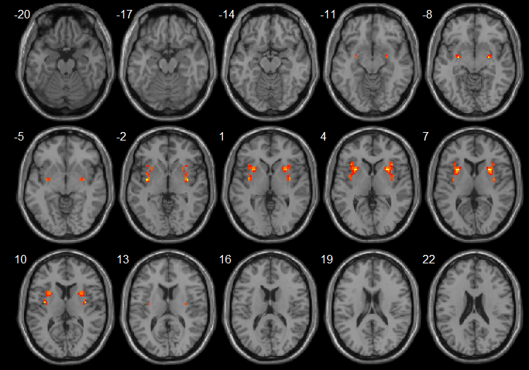
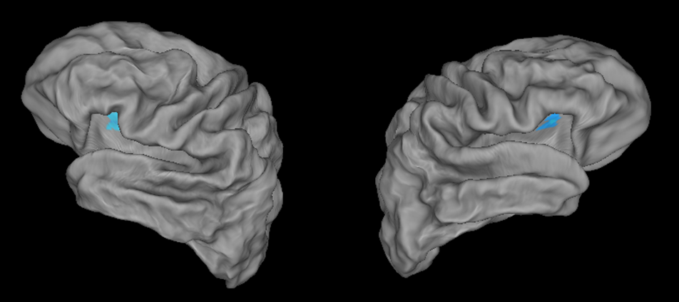
According to the definition, VMHC maps exhibited a bilateral symmetric pattern. The RLS group showed increased VMHC in the amygdala, putamen and insula, as compared to normal controls. Significantly different regions were overlaid on the standard T1 template of a single subject (Fig. 1) and the fiducial surface from Caret (http://brainmap.wustl.edu/caret ) (Fig. 2).Discussion and Conclusion
In this preliminary study, we found increased VMHC regions in the amygdala, putamen and insula for the RLS group as compared to normal controls. Increased VMHC in the insular cortex and putamen might reflect their functions in perception and motor control. Increased VMHC in the amygdalae could be relevant to the disturbed sleep at night, considering its primary role in the processing of memory and emotional reactions. Further study requires more subjects and needs to correlate the changes of inter-hemispheric functional activity with other clinical and biological indicators.Acknowledgements
No acknowledgement found.References
1. Trenkwalder C, Paulus W and Walters AS. The restless legs syndrome. Lancet Neurol. 2005; 4: 465–475.
2. Zang Y, Jiang T, Lu Y, et al. Regional homogeneity approach to fMRI data analysis. NeuroImage. 2004;22(1):394– 400.
3. Zuo XN, Kelly C, Di Martino A, et al. Growing together and growing apart: regional and sex differences in the lifespan developmental trajectories of functional homotopy. J Neurosci. 2010;30: 15034-15043.
Figures