1700
Localization of the epileptogenic neural network by the resting state fMRI in patients with the temporal lobe epilepsy1Human and Animal Physiology, Taras Shevchenko National University of Kyiv, Kyiv, Ukraine, 2Radiology Department, Shupyk National Medical Academy of Postgraduate Education, Kyiv, Ukraine
Synopsis
Precise localization of the epileptogenic zone and its delineation from
Introduction
Epilepsy is a chronic neurological disorder characterised by the recurrent seizures and it occurs in over 0.5% of the world population. Temporal lobe epilepsy (TLE) accounts for more than a half of cases in the epilepsy cohort1. Usually, TLE exhibits recurrent seizures and has its epileptogenic zone. Hippocampus plays important role in seizure origin as it possesses a large number of epileptogenic potassium ion channels2. Controversial data are present about the TLE influence onto the neural network functional connectivity in the brain. One part of authors claim that TLE disrupt the functional connectivity, while others support the idea of connectivity increase, especially in the local temporal lobe network3,4. Drug resistant epilepsy surgical treatment is relatively widespread. Precise localization of the epileptogenic zone and its delineation from eloquent cortex, are crucial for successful surgery. We propose the analysis of macroscopic brain neural networks connectivity in the resting state and during the movement execution in patients with TLE for the possible epileptogenic macroscopic neural network localisation and motor cortex mapping.Methods
Group of 8 patients (4M, 4F, age 19-49 y.o.) was studied with the functional MRI. Temporal lobe epilepsy (TLE) was diagnosed by the neurologist with the EEG. We used 1.5T SIGNA EXCITE (GE, USA) for routine MRI, DWI (b=1000) with ADC maps and fMRI. Simple finger tapping task (6 min, 6 blocks of activation) and resting state data acquisition was used for the fMRI. EPI sequence was used for BOLD imaging (TR/TE=3000/71 ms, voxel 4x4x5 mm). FMRI data processing was carried out using GLM (FEAT) and ICA (MELODIC) based software from FSL (Oxford, GB).Results
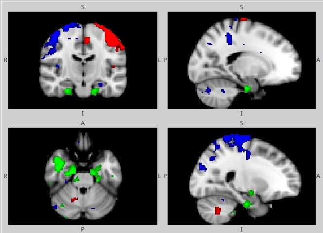
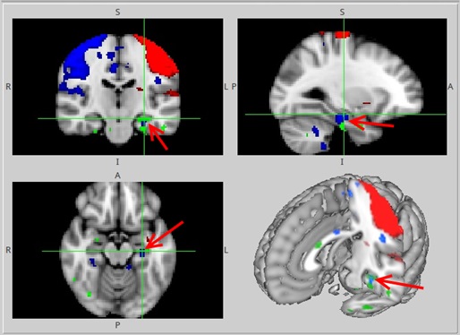
Functional MRI data analysis revealed activation of the intact motor neural network of the brain (Fig. 1, red). It consisted of the contralateral (left) primary sensorimotor cortices (pre- and postcentral gyri) supplementary motor area (medial dorsal superior frontal gyrus), ventral premotor area and ipsilateral (right) hemisphere of cerebellum. Deactivation of the left hippocampus was found during the movement execution. Resting state fMRI data analysis revealed increased functional connectivity of the anterior pole of the inferior temporal gyrus, posterior pole of the middle temporal gyrus, hippocampus and parahippocampal gyrus of the left hemisphere. Spectral analysis of the BOLD signal fluctuations revealed the main frequency of 0.001 Hz. It excludes the possibility of the artificial origin (heartbeat, breath movements) of the respective independent component. This independent component potentially depicts the epileptogenic neural network as it is topographically coincides with the localisation of the EEG epileptiform activity. Here we propose an data-driven method utilizing resting state fMRI data. Our results show the concordant topography of the resting state network and the EEG epileptiform activity. Currently fMRI, and especially correlated or simultaneously recorded with EEG is promising technique with high sensitivity and specificity for detection of epileptic zones5.Conclusion
To conclude, according to our data, resting state functional MRI could potentially depict the epileptogenic neural network in TLE which includes regions of temporal lobe and hippocampus. Also left hippocampus was shown to deactivate during the motor task execution. Motor network was shown to remain unaffected in TLE during interictal period.Acknowledgements
References
1. Zhang C.H., Sha Z., Mundahl J., et al., Thalamocortical relationship in epileptic patients with generalized spike and wave discharges — A multimodal neuroimaging study, NeuroImage: Clinical, Vol. 9, 2015, P.117-127,
2. Kues W. A. , Wu?der F. “Heterogenous expression pattems ofmammalian potassium channel genes in developing and adult rat brain” Eur. J. Neurosci., No. 4., 1296-1308, (1992).
3. Rajpoot K, Riaz A, Majeed W, Rajpoot N (2015) Functional Connectivity Alterations in Epilepsyfrom Resting-State Functional MRI. PLoS ONE 10(8):e0134944. doi:10.1371/journal.pone.0134944
4. Wurina, Zang YF, Zhao SG. Resting-state fMRI studies in epilepsy. Neurosci Bull. 2012 Aug;28(4):449-55.
5. Preti M. G. et al. "Epileptic network activity revealed by dynamic functional connectivity in simultaneous EEG-fMRI." 2014 IEEE 11th International Symposium on Biomedical Imaging (ISBI). IEEE, 2014.
Figures