1699
Impaired interactions of large-scale networks predict relapse behavior in heroin-dependent individuals1Tangdu Hospital, Fourth Military Medical University, Xi'an, People's Republic of China
Synopsis
Coupling of large-scale brain networks may underlie cognitive dysfunction in psychiatric disorders including addiction. However, whether the deficit of interactions among large-scale brain networks is associated with relapse behavior of heroin addiction remains unknown. This is the first neuroimaging study to assess coupling of large-scale networks that predict relapse in heroin addiction. In this study, we utilized a resting-state functional connectivity method of functional magnetic resonance imaging and found that abnormally higher functional connectivity between the SN and DMN, and lower functional connectivity between the left ECN and DMN were associated with the relapse behavior in treated heroin-dependent patients.
Purpose
Interactions of large-scale brain networks may underlie cognitive dysfunction in psychiatric disorders including addiction1. However, whether the deficit of interactions among large-scale brain networks is associated with relapse behavior of heroin addiction remains unknown. To the best of our knowledge, this is the first neuroimaging study to assess coupling of large-scale networks (salience, default mode and executive control) that predict relapse in heroin addiction.Methods
Fifty heroin-dependent patients and 20 healthy subjects attended a prospective resting-state functional magnetic resonance imaging study. The salience, default mode and executive control networks were identified by independent component analysis with Gift software and 9 core spherical region of interest within these 3 networks. The authors compared the functional connectivity among healthy controls, the relapsed and early remission patients defined based on urine tests during a 3-month follow-up. The authors also examined how the coupling of large-scale networks related to relapse behavior among heroin-dependent patients.Results
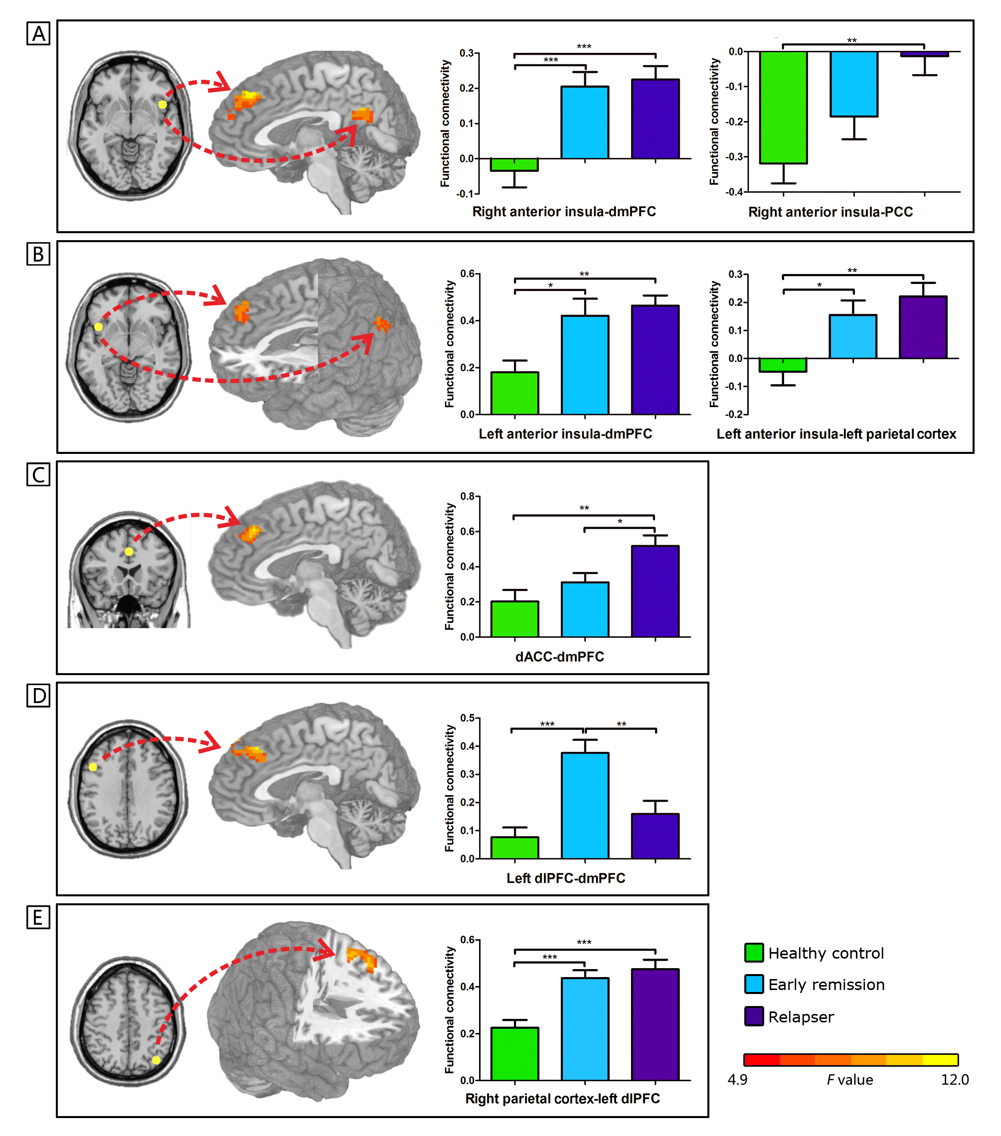
Twenty-six and 24 heroin-dependents were defined as relapsers and early remission patients respectively. The functional connectivity between nodes of salience and default mode networks is generally higher in the relapsers and early remission patients relative to healthy controls. The relapsers demonstrated higher connectivity between dorsal anterior cingulate cortex (a node of salience network) and dorsomedial prefrontal cortex (included in default mode network) relative to the early remission group. The relapser and healthy groups demonstrated lower connectivity between the left dorsolateral prefrontal cortex (a node of left executive network) and dorsomedial prefrontal cortex relative to the early remission group, respectively. (Figure 1) Across the heroin-dependent patients, the numbers of positive urine screen during 3 months follow-up positively correlated with the coupling between the dorsal anterior cingulate cortex and dorsomedial prefrontal cortex (r2 = 0.164, Pcorrected = 0.028), but negatively correlated with the coupling between the left dorsolateral prefrontal cortex and dorsomedial prefrontal cortex (r2 = 0.157, Pcorrected = 0.028).Conclusion
Higher coupling between the salience and default mode networks and lower coupling between the left executive and default mode networks predict relapse behavior. This findings may shed light on the development of new treatment for heroin addiction.Acknowledgements
This work was supported by grants from the National Natural Science Foundation of China (No. 81201081, 81371532, 81471648, 81401393, 81071142 and 81071143) and the grant from Technology Innovation Development Foundation of Tangdu Hospital (No. 2013LCYJ003).References
1. Morie KP, Garavan H, Bell RP, De Sanctis P, Krakowski MI, Foxe JJ. Intact inhibitory control processes in abstinent drug abusers (II): a high-density electrical mapping study in former cocaine and heroin addicts. Neuropharmacology. 2014; 82: 151-60.Figures