1685
Growing Apart or Growing Together: a Novel “Developing Triple Network” Hypothesis for Baby Connectome Study1Department of Radiology and BRIC, University of North Carolina at Chapel Hill, Chapel Hill, NC, United States, 2Department of Biomedical Engineering and BRIC, University of North Carolina at Chapel Hill, Chapel Hill, NC, United States, 3Department of Brain and Cognitive Engineering, Korea University, Seoul, Korea, Republic of
Synopsis
For baby connectome study, we propose a “developing triple network” model based on our findings from precious longitudinal infant resting-state fMRI data, which extends previous “triple network model” to 0-2 years old, a pivotal period which we have little knowledge on. Our developmental model provides future study with key information on early functional connectivity. We found medial prefrontal area is growing apart and its long-range FC is growing together at different speeds in the first 2 years. We, for the first time, found a novel early developmental pattern with both local and long-range FC increases at 6-9 months but decreases afterwards, indicating pivotal neural pruning mechanism from system neuroscience view point.
Introduction
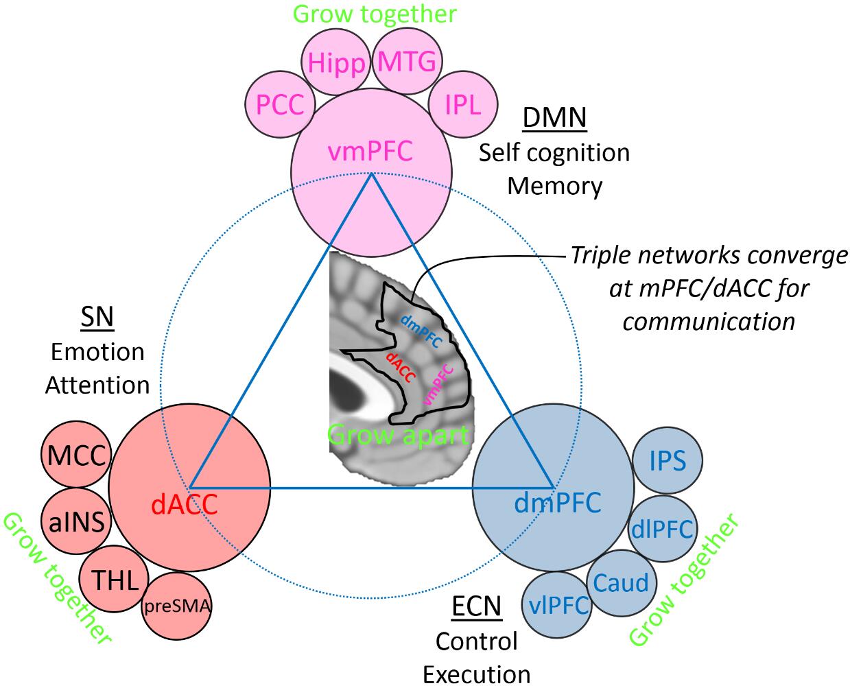
Infancy, a critical and important stage in human brain development, is not yet well-studied. Although we have gained abundant acknowledgement on brain connectome between childhood and aging periods, we are still in lack of understanding “baby brain connectome”. Non-invasive resting-state functional MRI (rs-fMRI) provides an unprecedented opportunity for this study. Understanding developing trajectory in this pivotal period is immensely important since any subtle change could cause tremendous consequences at adolescence. In adults, we have known that different brain functional connectivity (FC) networks carry out different perceptive and cognitive functions and interact with each other. Recently, an intriguing “triple-network” model was proposed based on the fact that many psychiatric and neurologic diseases are involved in this common structures [1]. In this model, default mode network (DMN), executive control network (ECN) and salience network (SN) interweave together, with the SN controlling attention re-allocation to support outward executive and inward self-referential activities. In this study, we focus on the triple networks and investigate its development during infancy, because detection of any subtle abnormality within this triangular relationship as early as possible is important to prevent severe late consequences. We noticed that, although the three networks involve spatially remote regions, they converge at a relatively small region encompassing two heavily connected areas, the medial prefrontal cortex (mPFC) and the dorsal anterior cingulate cortex (dACC). We hypothesize that this region will grow apart while each functional network will grow together on different time (Fig. 1).Methods
We collected 5-min rs-fMRI data multiple times for each infant during sleep at their 1, 3, 6, 9, 12, 18 and 24 months. Due to data drop off, we have total sample sizes of 33, 29, 31, 30, 35, 26 and 22 at each time point. Data preprocessing was performed using FMRIBs Software Libraries according to previous studies [2]. Of note, data at different time points were firstly aligned within subjects and then registered across different subjects into standard space using our 4D registration algorithm. Region of interest (ROI) was chosen based on Automated Anatomical Labeling atlas, including bilateral superior medial frontal areas and anterior cingulate cortex. At each time point, all infants’ data were fed into group independent component analysis to conduct functional parcellation of the ROI to investigate whether this ROI will grow apart. The derived sub-regions at 24 months were used as seed regions to further conduct whole-brain FC at each time point to investigate whether the regions within each functional network will grow together. Within-ROI FC between each pair of the sub-regions was calculated at each time points. Whole-brain FC of each pair of 200 brain regions was also investigated.Results
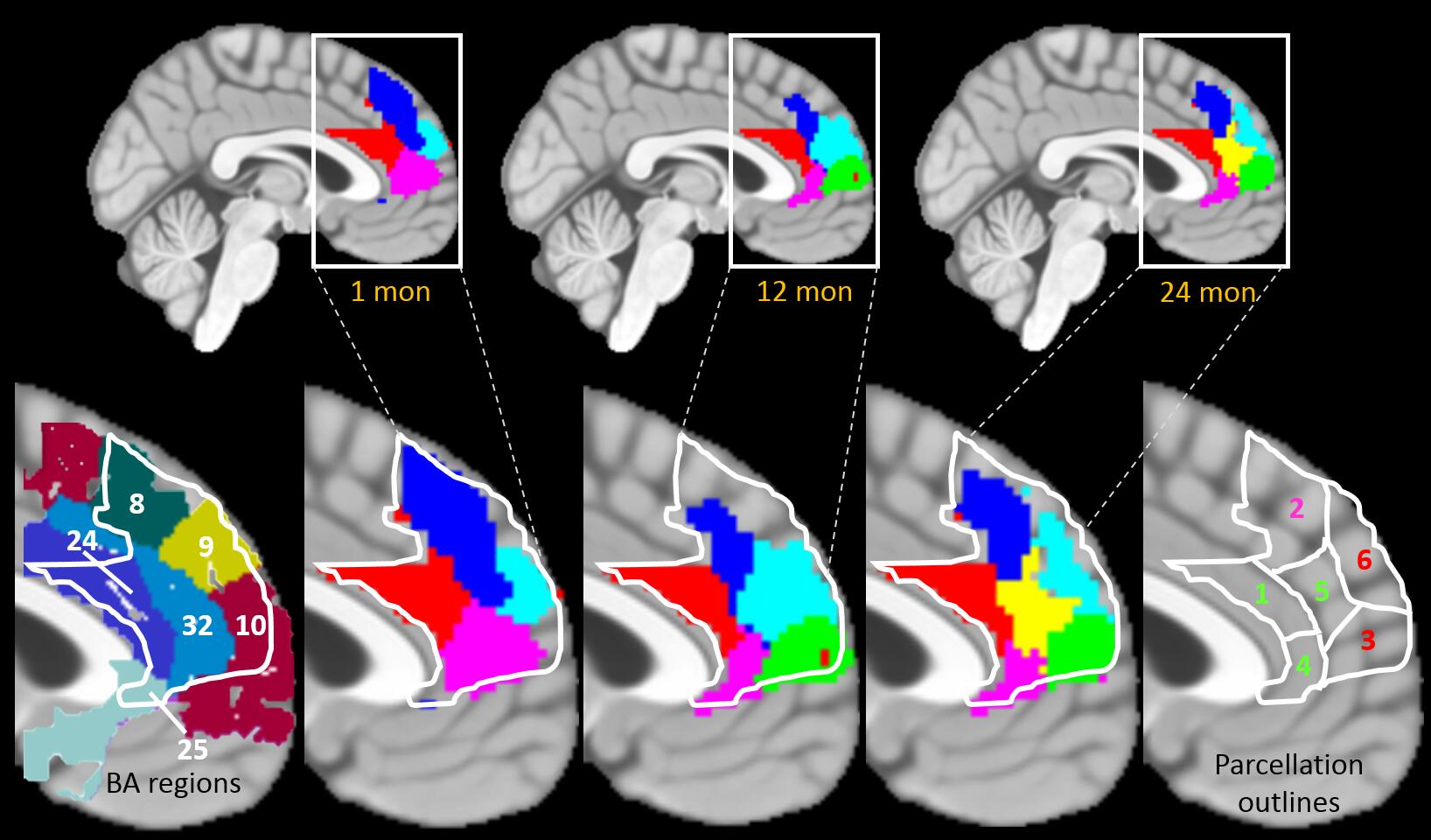
We found in mPFC/dACC, brain grows apart. Several sub-regions merge together in newborns but well separate after 2 years (Fig. 2). New sub-regions emerge: from temporal aspect, a yellow-colored sub-region in the center of the mPFC appears after 24 months; from spatial aspect, this sub-region sits in between different networks (red, blue and green areas in Fig. 2 represent the medial frontal parts of the SN, ECN and DMN, respectively).
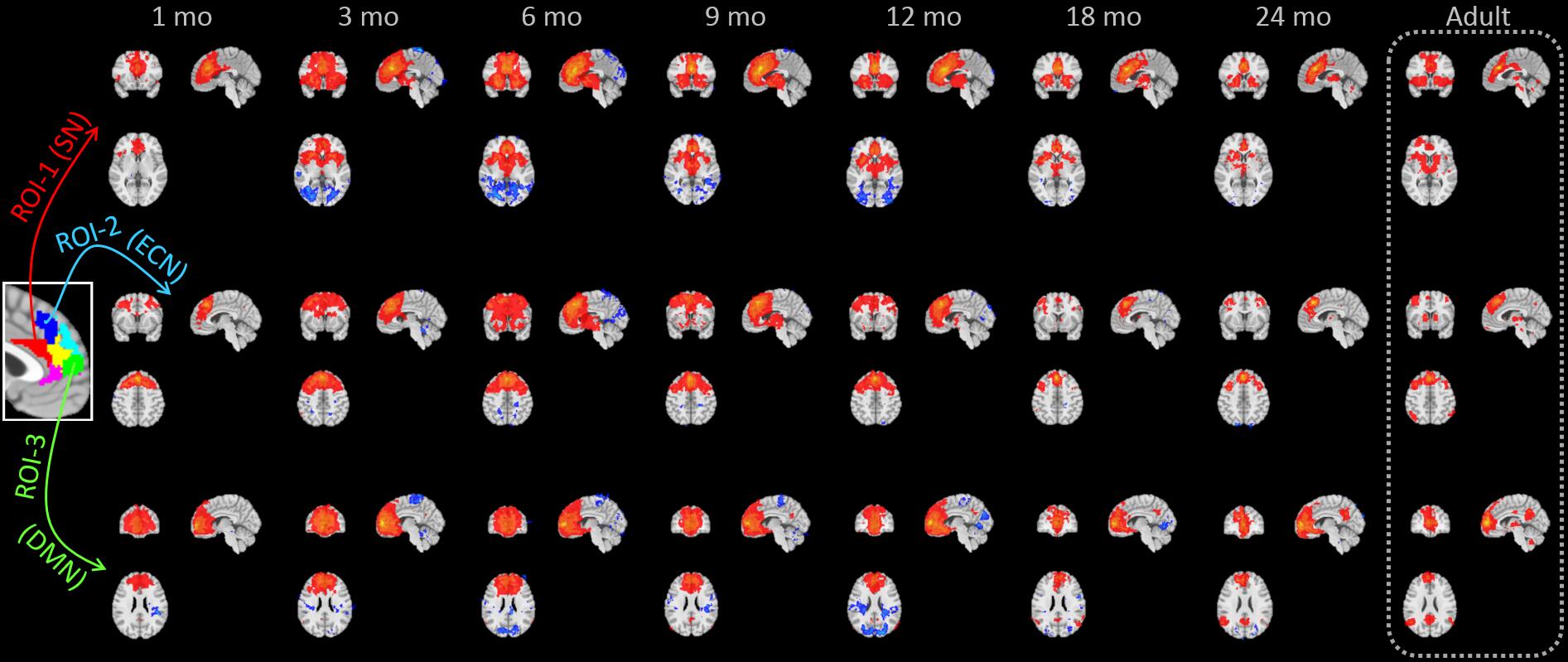
As shown in Fig. 3, different sub-regions gradually connect while developing (growing together). At 1 month, they hardly have any connections outside frontal areas and have their FC highly overlapped. At 2 years old, they get in touch with different networks, similar as the FC patterns of adults. Interestingly, their outward FCs development follows different temporal evolution courses with the maturing order: SN, ECN and DMN.
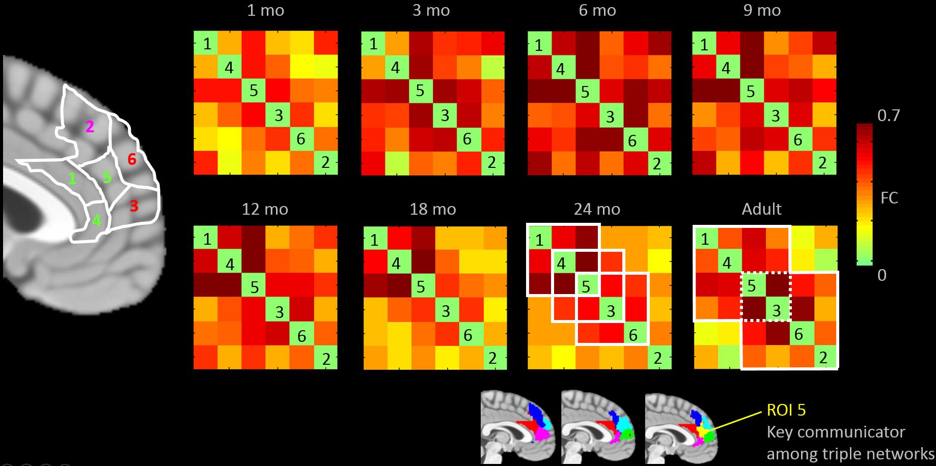
In Fig. 4, at 24 months, sub-region 5 (the yellow region) grows mature and connects with all three networks via a triple overlap in the connectivity matrix, indicating that information collected from the three networks merges and interacts in this sub-region.
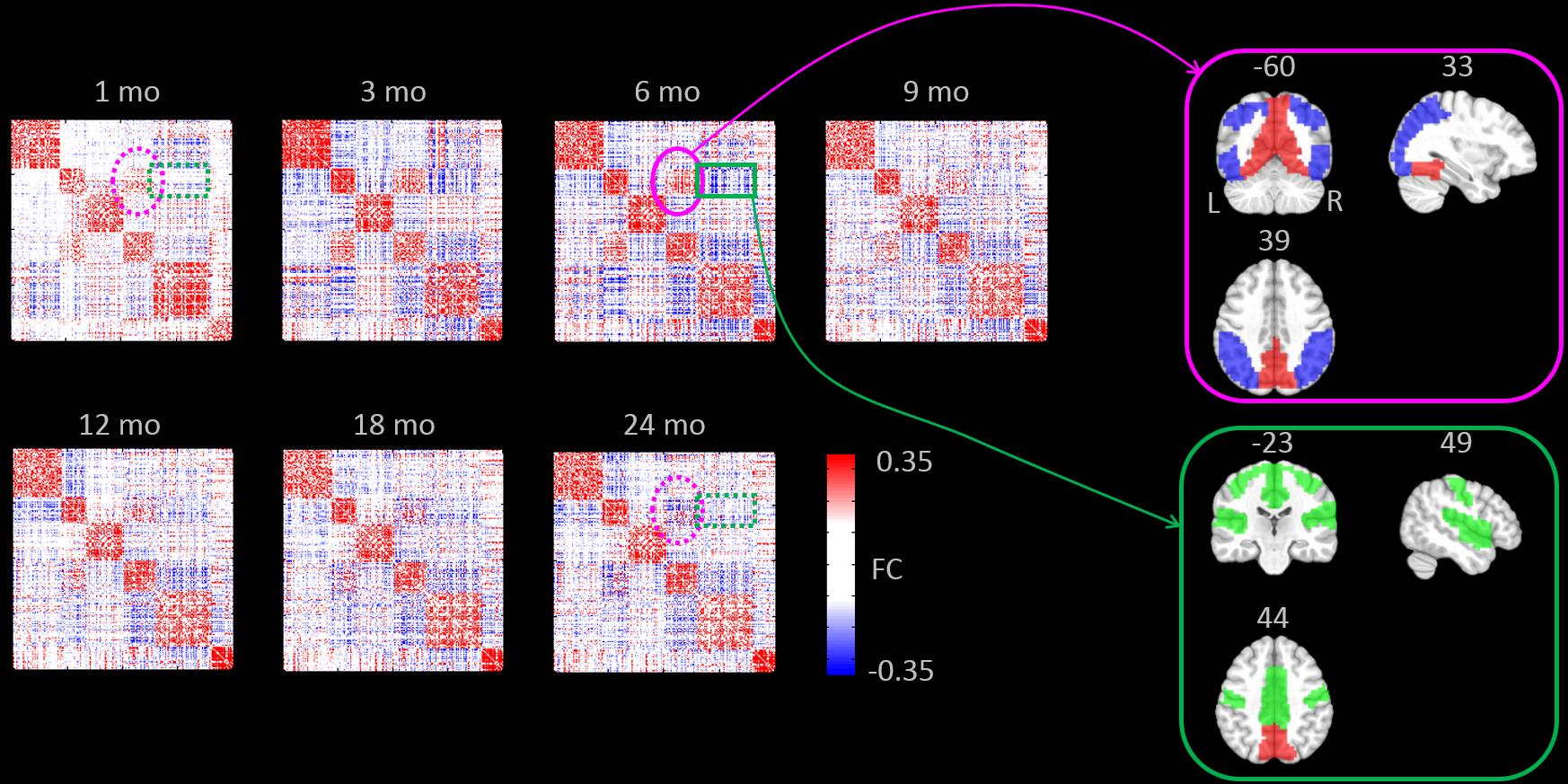
From both local, within-ROI FCs (Fig. 4) and global, whole-brain FCs (Fig. 5), we found a significant "sparse-dense-sparse" developmental pattern. The densest FCs occur between 6-9 months; at this period, two visual networks are strongly connected, while the primary visual and primary sensorimotor networks are strongly anti-correlated. This indicates visual information integration and multisensory suppression is at peak at 6-9 months.
Conclusion
Our hypothesis that mPFC/dACC area grows apart and its long-range FC within each network grows together at different speeds in the first 2 years is proven. We propose a “developing triple network” model. Importantly, we found a novel early developmental pattern, where both local and long-range FCs increase at 6-9 months and decrease afterwards. This finding, for the first time, indicate pivotal neural pruning mechanism from system neuroscience point of view.Acknowledgements
No acknowledgement found.References
[1] Menon V. Large-scale brain networks and psychopathology: a unifying triple network model. Trends Cogn. Sci. 2011; 15: 483–506.
[2] Gao W, Alcauter S, et al. Functional Network Development During the First Year: Relative Sequence and Socioeconomic Correlations. 2015; 25: 2919–2928.
Figures