1680
Effect of Repeated Injection of Iodixanol on Rrenal Function in Healthy Wistar Rats by Using Functional MRI1Department of Radiology, First Affiliated Hospital of China Medical University, Liaoning, People's Republic of China, 2GE Healthcare, MR Research China, Beijing, Beijing, People's Republic of China
Synopsis
In clinical practice, for complex, recurrent disease, the same patient may need to use contrast agents repeatedly. However, the administration of multiple use of contrast-medium within a short period of time may put patients at risk1 .So, the aim of this work is to examine differences in rats’ potential kidney injury provoked by iodixanol with respect to different intervals and different times. By using fMRI we observed whether repeated intravenous injection of iodixanol at different time intervals could lead to aggravate kidney damage and then find the shortest time interval. Simultaneously, monitoring kidney damage by sNGAL and detecting the cause of renal injury by immunohistochemistry were also performed.
Purpose
To determine the appropriate time interval between repeated contrast-medium administrations by comparing the effects of repeated intravenous injections of iodixanol at different time intervals, and to investigate the injury location and causes of renal damage in rats (acute model).Material and Methods
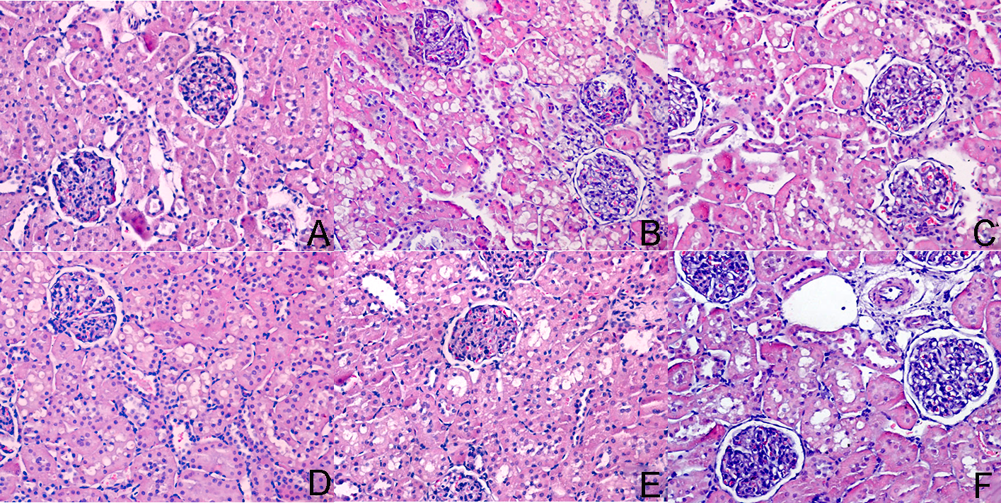
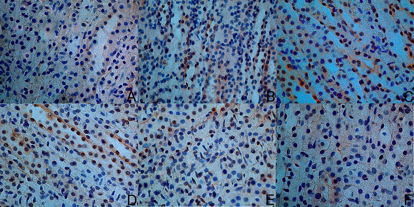
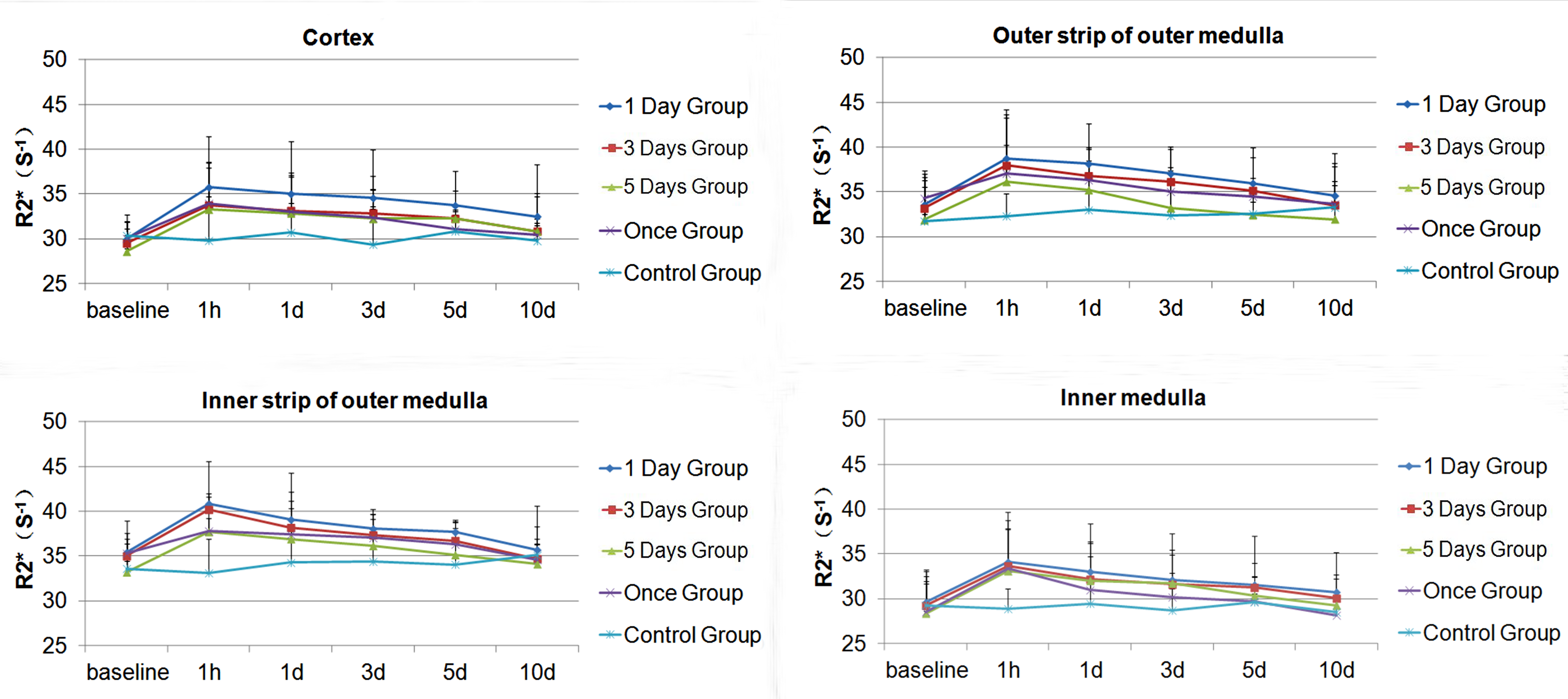
The animal study protocol was approved by the institutional ethics committee of China Medical University. According to the number of iodixanol injection, Wistar male rats 300+/-20g (n=83) were randomly divided into Once group (n=17), Twice group (n=51) and Control group (n=15). The Twice group was subdivided into 3 cohorts according to the interval between the first and second injections of iodixanol (1 day, 3 days, 5 days). Intravenous contrast was administrated at 4g iodine/kg of body weight. BOLD-fMRI was performed at 1 hour, 1 day, 3 days, 5 days, 10 days after injecting saline or iodixanol, and three rats were sacrificed at each time point. Each kidney was divided into four regions following correlation with corresponding histological sections according to Li et al2 (Figure 1). They were corresponding to cortex (CO), outer strip of outer medulla (OSOM), inner strip of outer medulla (ISOM) and inner medulla (IM). The expression of hypoxia inducible factor (HIF-1α) in renal tissue was detected by immunohistochemistry. Renal injury was confirmed using urinary neutrophil gelatinase-associated lipocalin (uNGAL) measurements. The SPSS17.0 statistical software was used for data analysis, P value less than 0.05 was considered statistically significant. Rank sum tests were used to obtain non-normal distributions or heterogeneity of variance. Statistical analysis was performed by using one-way analysis of variance with LSD and T-test.Results
Differences in BOLD-fMRI results among the different groups were observed in all 4 renal regions (Figure 2).ISOM showed the most pronounced changes of all 4 renal regions. Iodixanol increased the R2* values to the maximum levels at 1h (Table 1), which is in line with several studies3-5.For R2* values of ISOM at the 1-hour time point, the 1-day and 3-days groups were significantly different from the Once group (P=0.015, P=0,039, respectively), but the 5-days group was not statistically significant (P>0.05). These results indicated that short-term repeated injection of iodixanol is the risk factors of CIAKI and a 5-days
interval is the optimal time interval. In the 1-day group, R2* values increased significantly over time. Compared with baseline, R2* values were significantly higher in CO in the 10-day group (P=0.025), the pathological tissue also showed some intracytoplasmic vacuoles in the renal tubular epithelia (Figure 3). What’s more, a complete or near-complete recovery of baseline renal function occurs within 10 days in the 3-days group, meanwhile, data from the 5-days group were close to baseline at day 5. After repeated injection of iodixanol, hypoxia inducible factor HIF-1α were transiently upregulated at 1 hour, and the expression of HIF-1α was the most significant in ISOM area. HIF immunostaining was confined to a short period of time, within 5 days after the induction of the hypoxic insult in the 1-day group (Figure 4). No statistically significant change was found for urinary neutrophil gelatinase–associated lipocalin (uNGAL) after iodixanol administration within 5 days for neither the Once or Twice group.
Conclusion
Repeated injection of iodixanol contrast agent within a short time can cause significant renal damage and it is more likely to cause acute kidney injury, and repeated occurrence of acute kidney injury can lead to chronic kidney injury. The shorter the interval between repeated injections, the more severe the kidney damage in rats. And the resulted renal damage and recovery time last longer. The effect of repeated injections of iodine contrast agent on renal damage in rats will gradually disappear after more than 5 days.Acknowledgements
No acknowledgement found.References
1. Stacul F, et al.[J]. European radiology. 2011,21(12):2527-41.
2. Li LP, et al. Investigative radiology. 2014,49(6):403-10.
3. Wang YC, et al. Investigative radiology. 2014,49(11):699-706.
4. Li LP, et al. J Magn Reson Imaging. 2012,36(5):1162-7.
5. Lenhard DC, et al. Invest Radiol. 2013;48(4):175-82.
Figures

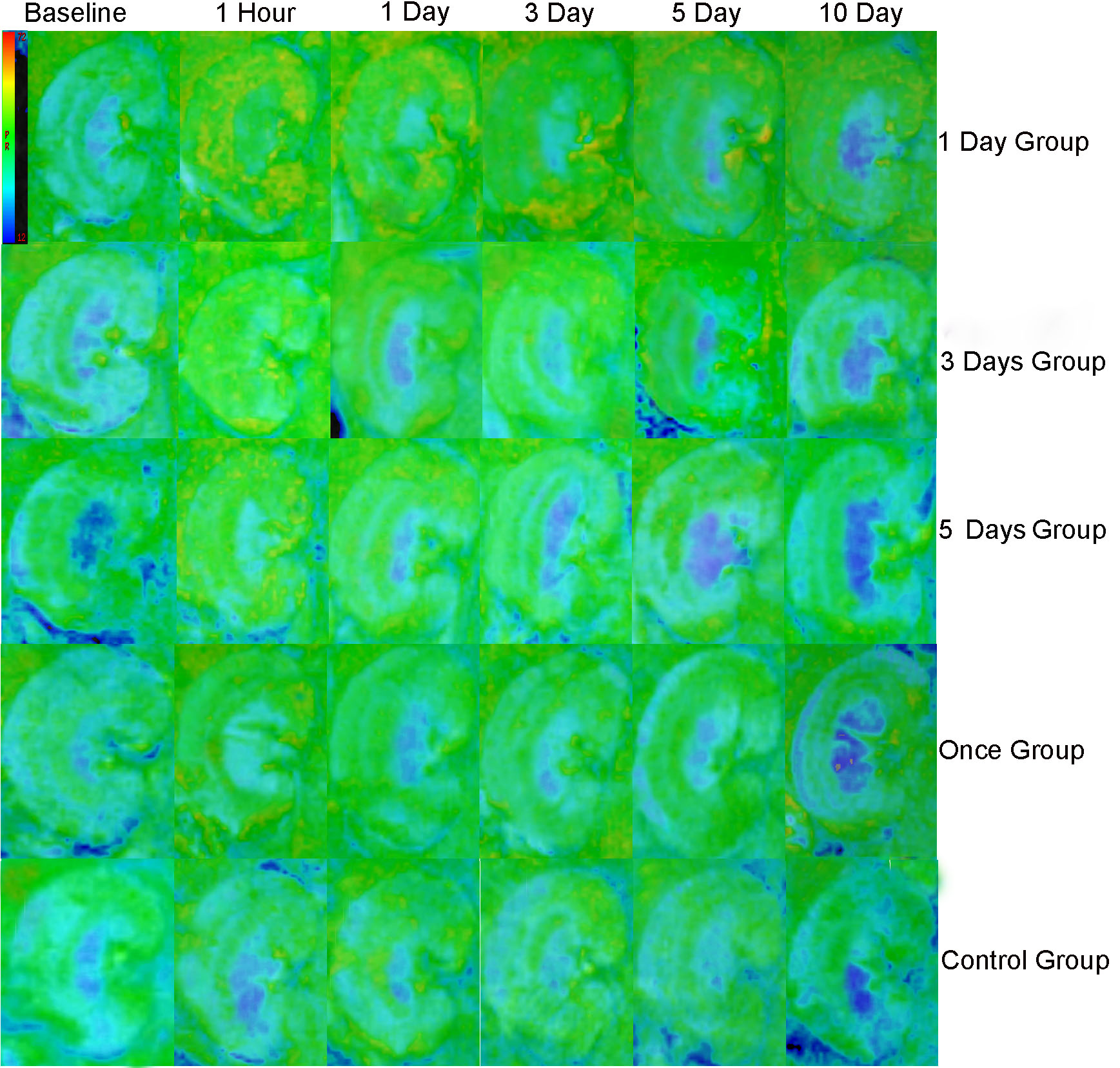
Figure.2 Representative R2* maps were obtained at baseline, 1 hour,1 day,3 day, 5 day and 10 day in the iodixanol group.1 Day Group,3 Days Group and 5 Days were injected repeatedly at different time intervals. Once Group’s rats were injected with iodixanol only once.15 rats were sacrificed as control group with injection of saline. Note the cortex in 1 Day Group showed persistent elevation of R2* for 10 days. The intensity of the ISOM was greater than that in the CO, OSOM and IM, which implies lower oxygenation of ISOM than the other regions.