1677
Dose-dependent effects of prolonged isoflurane administration on resting cerebral blood flow and functional connectivity: a preliminary study in rhesus monkeys1Yerkes Imaging Center, Yerkes National Primate Research Center, Emory University, Atlanta, GA, United States, 2Division of Neuropharmacology and Neurologic Diseases, Yerkes National Primate, Emory University
Synopsis
Isoflurane is widely used in biomedical research
with the prolonged duration of administration up to several hours. However, the
manner in which neurophysiology and functional connectivity are affected by the
length of anesthesia remains poorly understood. In the present study, cerebral
blood flow (CBF) and default model network (DMN) were examined using arterial
spin-labeling perfusion and resting state functional MRI techniques. The functional connectivity in the
dominant DMN decreased substantially during 4-hour administration of isoflurane
at any given dosage. CBF in most brain
regions decreased at the low dose (0.89 %), but did not change markedly when higher doses of isoflurane (1.05 %,
1.19 %) were administrated. The study revealed dose-dependent effects of
isoflurane on brain functionality and regional CBF during prolonged anesthesia
administration of isoflurane, suggesting those effects should be considered in
the experimental design or the interpretation of the outcome of related
neuroimaging studies using anesthetized animals or humans.
Introduction
Prior studies have demonstrated that prolonged administration of isoflurane could alter the regional CBF in anesthetized brains and might be associated with the post-anesthesia cognitive impairment [1-10]. In the present study, adult rhesus monkeys were used to examine the dose-dependent effects of isoflurane during the 4-hour administration on CBF and brain connectivity by using the continuous arterial spin-labeling (CASL) perfusion MRI and resting-state functional MRI (rsfMRI) techniques.Methods
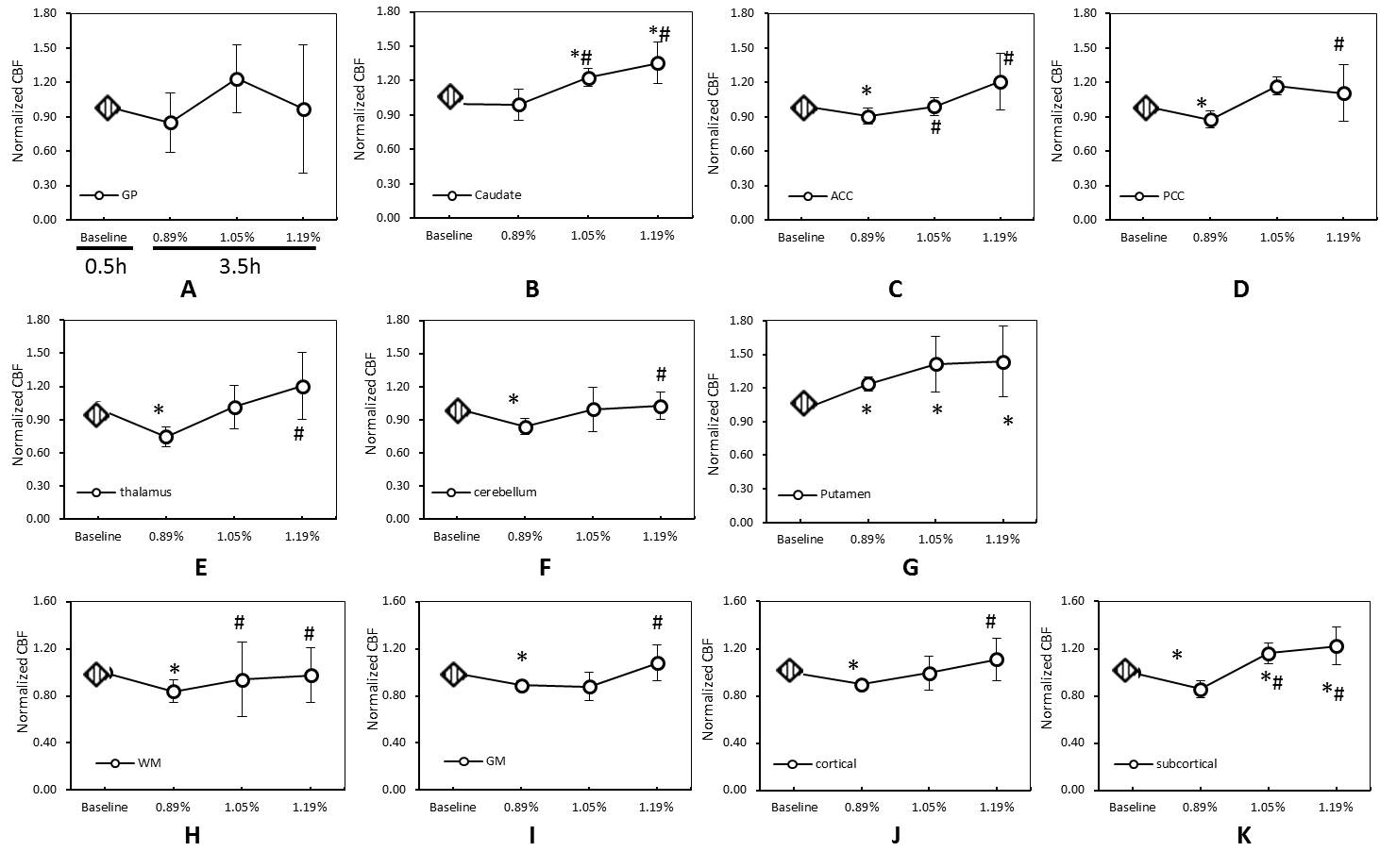
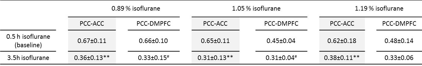
Five rhesus monkeys (7-11 years old) were anesthetized with isoflurane mixed with 100% oxygen for approximately 4 hours. O2 saturation, blood pressure, heart rate, respiration rate, body temperature and PaCO2 were monitored continuously and maintained in normal ranges. Three dosages of isoflurane (0.89 ± 0.03 % (~0.8 MAC), 1.05 ± 0.03 % (~0.9 MAC), and 1.19 ± 0.09 % (~1.1 MAC), mean ± standard deviation (SD), end-tidal concentration) were used. A gradient echo EPI sequence (TR/TE=2190 ms/25ms, 430 volumes) were used to acquire rsfMRI data using a Siemens 3T TIM Trio with an 8-channel Tx/Rx volume coil. CBF was acquired using a pCASL sequence with TR/TE=3830ms/21ms, voxel size = 1.5×1.5×1.5mm3, 6 repetitions. The fMRI and CBF data were collected firstly at the 0.5-hour time point (tp0.5) and rescanned at the 3.5-hour time point (tp3.5) during each scan session. CBF data analyses were performed using home-built Matlab scripts and the Stimulate software [11]. The bilateral caudate, putamen, globus pallidus (GP), anterior cingulate cortex (ACC), posterior cingulate cortex (PCC), thalamus, cerebellum, white matter (WM), grey matter (GM), cortical and subcortical cortex were selected as regions of interest (ROIs). Paired t -test was performed to analyze the CBF differences statistically. CBF at tp3.5 was normalized to its baseline (tp0.5) to minimize the inter-subject variation. rsfMRI data were processed by distortion correction, slice timing correction, rigid body registration, regressing out signal in WM and cerebrospinal fluid time series, temporal filtering with 0.009 Hz ~0.0237 Hz band-pass, spatial smooth by a Gaussian blur with 2.5-mm full width at half maximum using an AFNI script (http://afni.nimh.nih.gov) [12]. The ROIs included the whole PCC, ACC, and dorsal/media prefrontal cortex (DMPFC) and were selected using AFNI and the monkey brain atlas [13] with T1-weighted images as reference. The averaged time courses of rsfMRI signal in PCC were used to perform seed-based correlation analysis. The averaged z values were examined for statistical differences by using SPSS 21.0. P-values less than 0.05 were considered statistically significant.
Results
The dose-dependent effects of 4-hour administration isoflurane on CBF are illustrated (Fig.1) for each ROI. CBF in ACC, PCC, thalamus, cerebellum, GM, WM, cortical and subcortical decreased significantly 3.5 hours post anesthesia at 0.89 % isoflurane (Fig 1 C, D, E, F, H, I, J, K), but remained almost unchanged at 1.05 % or 1.19 % isoflurane. However, CBF in putamen and caudate, subcortical increased significantly at the end time point with higher doses of isoflurane compared to the baseline (Fig. 1 B, G). Similar suppression of functional connectivity (FC) in the default model network was observed at every dose of isoflurane (Table 1). The correlation degree (z score) of PCC with either DMPFC or ACC was obviously decreased with any dosage (p < 0.01) (Table 1).Discussion and conclusion
The present study has demonstrated evident dose-dependent effects of 4-hour isoflurane on CBF and FC of adult rhesus monkeys. The CBF result with higher dose of isoflurane was in good agreement with Kuroda et al’s reports in which 1.5 MAC [14, 15] and 1.0 MAC of isoflurane [16] were applied in humans for 3 and 4 hours, respectively. In addition, the CBF increase in subcortical regions of animals with 1.05 % or higher isoflurane (Fig 1K) agree partially with McPherson et al ’s study in which CBF in forebrain and hindbrain regions of NHPs increased with 1 MAC (1.0 %) isoflurane for 4 hours [10]. Prior studies have demonstrated that isoflurane altered the CBF in the brain and suppressed neural activity in a dose-dependent manner [11, 17]. As shown in our data, FC was reduced substantially after 3 hour scan at any given dose of isoflurane. The association of FC and CBF changes was seen at low dose of isoflurane but disappeared at high dose, suggesting the coupling between CBF and functional activity is strongly affected by the isoflurane dosage and duration, further investigation is needed in the future studies.In conclusion, there exist evident dose-dependent effects of isoflurane on neurophysiology in long-term anesthetized rhesus monkeys. The dosage and duration effects of anesthesia should be considered in the experimental design of fMRI and data interpretation with prolonged administration of isoflurane.
Acknowledgements
The project was funded by the National Center for Research Resources (P51RR000165) and is currently supported by the Office of Research Infrastructure Programs (OD P51OD011132).References
[1] Zhang, B., et al. Anesth Analg (2012); [2] Xie, Z., et al., J Gerontol A Biol Sci Med Sci (2006); [3] Wang, H., Ann Fr Anesth Reanim (2012); [4] Brambrink, A.M., et al., Ann Neurol (2012); [5] Birdsill, A.C., et al.,Obesity (Silver Spring) (2013); [6] Hirsch, C., et al. Dement Geriatr Cogn Disord (1997); [7] Poels, M.M., et al., J Cereb Blood Flow Metab (2008); [8] Jagust, W.J., et al. Brain (1992); [9] Roald, O.K., et al., Acta Anaesthesiol Scand (1989); [10] McPherson, R.W., Anesthesiology (1994); [11] Wu, W.C., et al., MRM (2007); [12] Chun-Xia Li., et al. ISMRM (2012); [13] Logothetis, K.S.S.a.N.K. A combined MRI and histology atlas of the rhesus monkey brain in stereotaxic coordinates. 2007(First edition); [14][15] Kuroda, Y et al., Anesthesiology (1996, 1997); [16] Kuroda, Y et al., J Anesth (2000); [17] Li, C.X., et al., Magn Reson Imaging (2014); [18] Smith, R.A et al., Biochim Biophys Acta (1981).Figures