1674
Hemodynamic and neuronal response in rat under Isoflurane-supplemented Medetomidine sedation assessed by simultaneous BOLD fMRI and Ca2+ recordings1Department of Clinical Radiology, University of Muenster, Muenster, Germany
Synopsis
fMRI in rodents has become a widely used technique with various applications in preclinical research. In this context, most studies are performed with anesthetized animals. Medetomidine evolves as a frequently used sedative but long term stability is limited -prolonged stability can be achieved by addition of Isoflurane. Several recent fMRI studies have used this combination. We evaluated the effects of this combination of anesthetics using a multimodal setup of BOLD fMRI and Ca2+ recordings. Even small addition of Isoflurane resulted in a significant decrease of neural (Ca2+) and hemodynamic (BOLD) responses to a distinct electrical stimulus.
Purpose
To quantify the impact of anesthesia consisting of a constant Medetomidine infusion and varying Isoflurane concentrations on fMRI and neural activity in rat, measured by simultaneous optical recordings with a genetically encoded Ca2+ sensor (GCaMP).Methods
BOLD
fMRI 20 rats (10 female Fischer, 5 male SD and 5 female SD) were investigated by fMRI, 3 rats additionally obtained simultaneous Ca2+ recordings. Animals were initially anesthetized using 5% Isoflurane. Electrodes were inserted into one forepaw and animals were transferred to a 9.4 T small animal MRI. Medetomidine infusion was started (0.04 mg/kg bolus, followed by infusion of 0.05 mg/kg/h) and Isoflurane was discontinued. Measurements began 40 min after Medetomidine infusion was started. The forepaw was electrically stimulated (5s stimulation (9Hz, 1mA), 25 s rest), repeatedly for 600 s. fMRI BOLD was performed using a single-shot GE-EPI (TR=1 s, TE= 18 ms, 600 repetitions, resolution 0.32 x 0.35 mm2, 9 slices, slice thickness 1.2 mm). Stimulation experiments were performed under stepwise increased and later decreased Isoflurane concentrations (seven steps: 0.0%, 0.2%, 0.4%, 0.7%, 1.0%, 1.25%, 1.5%). fMRI was repeated for each step, using the same stimulation paradigm, and allowing 10 minutes waiting time after concentration had been adjusted.
Ca2+ + BOLD
AAVs encoding GCaMP were injected into the somatosensory cortex of the forelimb (S1FL). After at least 4 weeks rest, animals were anesthetized with ~2% Isoflurane. A glass fiber (diameter 200 µm) was implanted in S1FL and was connected to a blue laser (488 nm) delivering excitation light. Using the same fiber, fluorescent light was detected and sampled with 2 kHz (fig. 1).1 Medetomidine infusion was started and Isoflurane was discontinued. The animal was transferred to the MRI for simultaneous BOLD + Ca2+ recordings. Anesthesia, stimulation and fMRI stayed the same. We additionally recorded Ca2+ responses to 3 Hz stimulation (1mA, 5 s stim.+ 25 s rest) for 300 s for each Isoflurane concentration. Animals subject to Ca2+ recordings were perfused transcardially and brains were removed for histological validation of GCaMP expression (fig. 4). fMRI analysis was performed with SPM 8. Using custom-written MATLAB scripts, a ROI (3x3 Voxel) was defined in the activated area and the amplitude of the BOLD timecourse was extracted. The amplitude the Ca2+ signal on stimulation was also analyzed and was reported with reference to baseline fluorescence (Δf/f). Onset and full width at half maximum was also analyzed.
Results
BOLD
Increasing Isoflurane concentrations gradually reduced the amplitude of the BOLD response. Addition of 0.2% Isoflurane reduced the amplitude by 27% (p=0.014), 0.7% Isoflurane resulted in a reduction of 62% (p<0.001). (fig. 3a)
Ca2+
Increasing Isoflurane concentrations gradually reduced the amplitude of detected calcium signal. Addition of 0.2% Isoflurane reduced the amplitude by 39% (p=0.038), 0.7% Isoflurane by 63% (p<0.001). Onset and full width at half maximum did not change significantly. For stimulation with 3 Hz, primary responses upon each stimulus were observed, regardless of the Isoflurane concentration (fig. 2a,d,g). For stimulation with 9 Hz, at high Isoflurane concentrations responses were observed only for the first pulses of the stimulus train (fig. 2b,e,h).
No significant differences between the results of test series with increasing (first half of experiment) vs decreasing (second half of experiment) Isoflurane concentrations were observed, nor between male vs female, nor between Fischer vs SD rats.
Discussion
Previous studies already stated that adding Isoflurane to Medetomidine sedation provides stable physiological conditions for prolonged fMRI studies. However, adding Isoflurane comes at the expense of losing signal in both fMRI (hemodynamic + neural signal) and Ca2+ recordings (neural signal).
The
combination of both anesthetics reduces the MAC of Isoflurane2 meaning that already little admixture
of Isoflurane has significant effects - the amplitude of the BOLD response is
decreased significantly. Isoflurane induces vasodilation of cerebral vessels3 In contrast, Medetomidine causes
vasoconstriction of both arteries and veins4, and attenuates cerebrovascular
dilation when combined with Isoflurane5. One could explain the effects observed
in BOLD with hemodynamic effects alone. Ca2+ recordings however indicate
that neural activity changes, too. This effect was not an effect of time
because while decreasing the Isoflurane concentration stepwise, both BOLD and Ca2+
amplitude recovered.
Conclusion
Even low doses of Isoflurane have big effects on hemodynamics and neural activity. However, our data suggests that addition of Isoflurane stabilize the animal’s physiology. Low doses of Isoflurane should provide a stable physiology while neural activity and hemodynamics is decreased on a tolerable level.Acknowledgements
No acknowledgement found.References
1. Schmid F, Wachsmuth L, Schwalm M, et al. Assessing sensory versus optogenetic network activation by combining (o)fMRI with optical Ca2+ recordings. Journal of cerebral blood flow and metabolism : official journal of the International Society of Cerebral Blood Flow and Metabolism 2015
2. Escobar A, Pypendop BH, Siao KT, Stanley SD, Ilkiw JE. Effect of dexmedetomidine on the minimum alveolar concentration of isoflurane in cats. Journal of veterinary pharmacology and therapeutics 2012; 35: 163–168
3. Iida H, Ohata H, Iida M, Watanabe Y, Dohi S. Isoflurane and Sevoflurane Induce Vasodilation of Cerebral Vessels via ATP-sensitive K+Channel Activation. Anesthesiology 1998; 89: 954–960
4. Fukuda M, Vazquez AL, Zong X, Kim SG. Effects of the alpha(2)-adrenergic receptor agonist dexmedetomidine on neural, vascular and BOLD fMRI responses in the somatosensory cortex. The European journal of neuroscience 2013; 37: 80–95
5. Ohata H, Iida H, Dohi S, Watanabe Y. Intravenous Dexmedetomidine Inhibits Cerebrovascular Dilation Induced by Isoflurane and Sevoflurane in Dogs. Anesthesia & Analgesia 1999; 89: 370–377
Figures
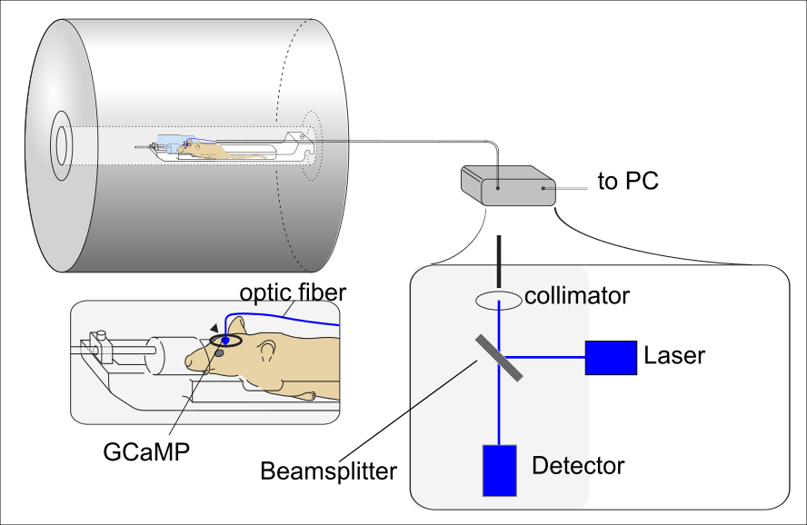
Figure 1
Experimental set up: The animal lies in the bore of a 9.4 T small animal MRI. A surface coil (d = 1 cm) is placed on S1FL, an optic fiber is led through the center of the coil for optic Ca2+ recordings. The fiber is connected to a blue laser (488 nm).
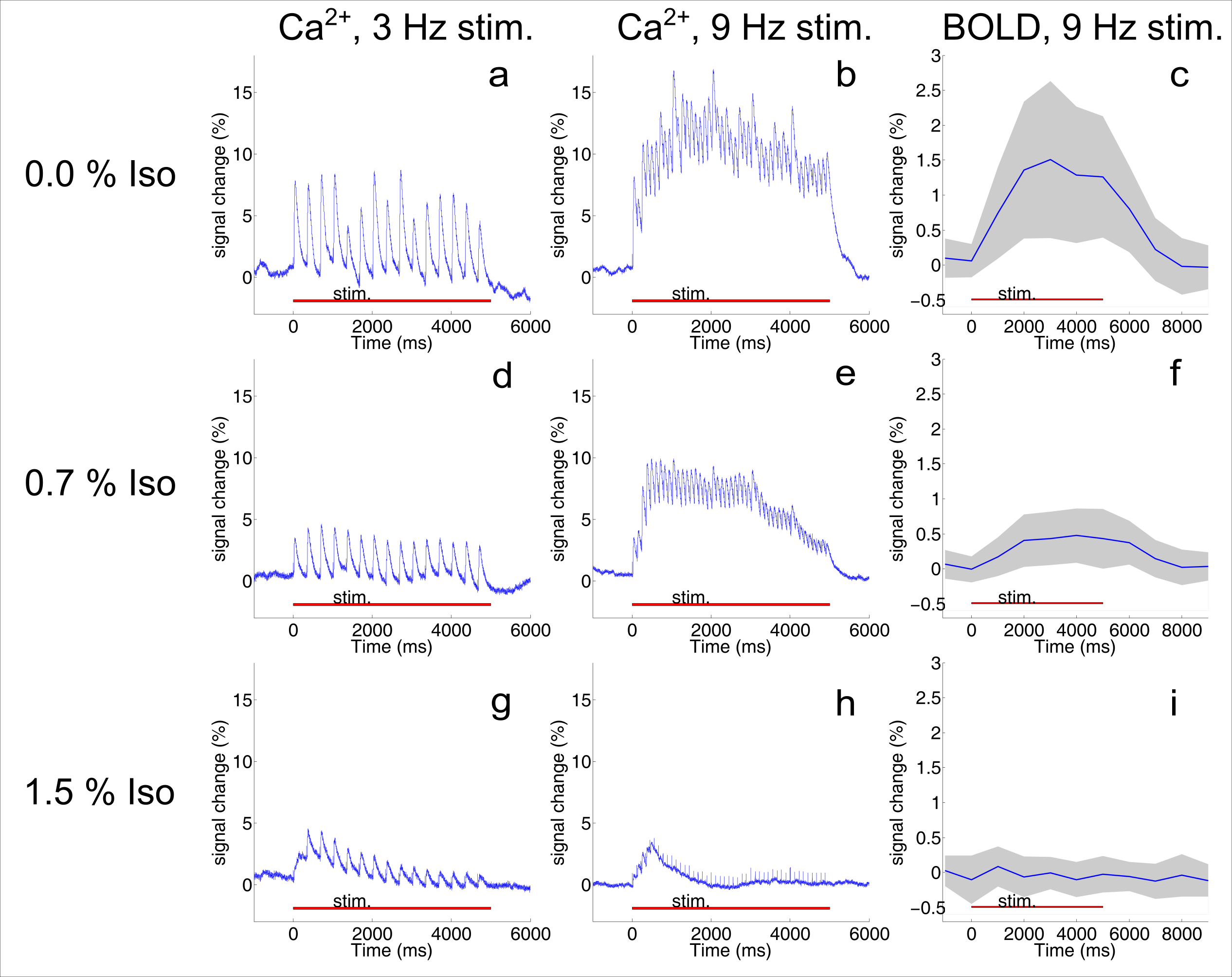
Figure 2
The first column shows the mean Ca2+ responses of one animal to stimulation with 3 Hz (averaged over 20 stimulation trains), the second column to stimulation with 9 Hz. Column 3 shows the mean BOLD response of all animals with the standard deviation (gray shading). Ca2+ recordings show primary responses corresponding to the frequency of the stimulus. The amplitude of the Ca2+ responses and of the BOLD response decreases with increasing isoflurane concentrations both at 3 Hz and 9 Hz.
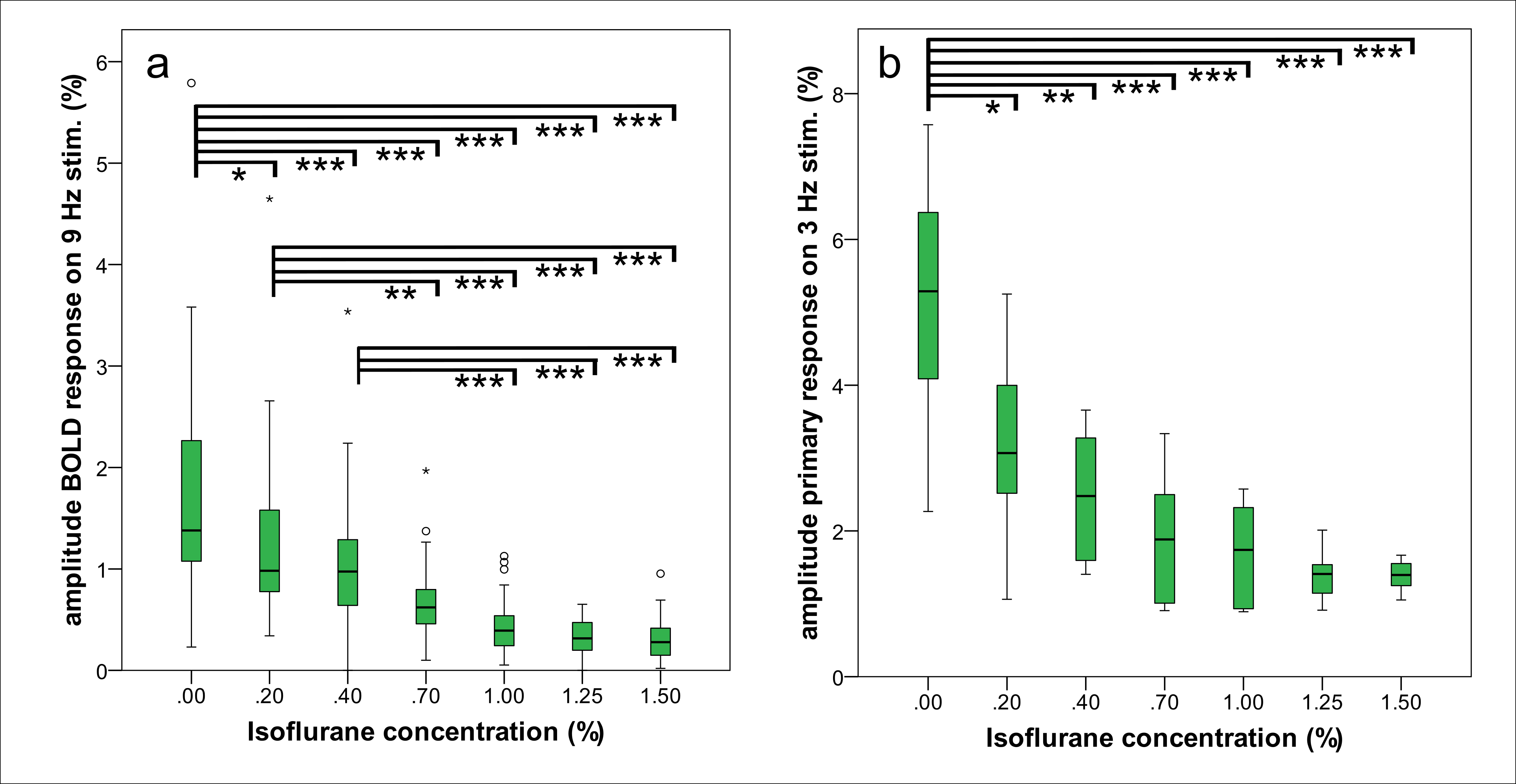
Figure 3
* = p <0.05
** = p <0.01
*** =p<0.001
a) Already addition of 0.2% Isoflurane to MED infusion reduced the amplitude of the BOLD response significantly. In total, 20 animals were subject to fMRI. Since there was no significant difference between test series with increasing and decreasing Isoflurane concentrations, data were combined (n=40).
b) Already addition of 0.2% Isoflurane to MED infusion reduced the amplitude of the Ca2+ signal. Equally to fMRI analysis, there was no significant difference between test series with increasing and decreasing Isoflurane concentrations, so data were combined (n=6).
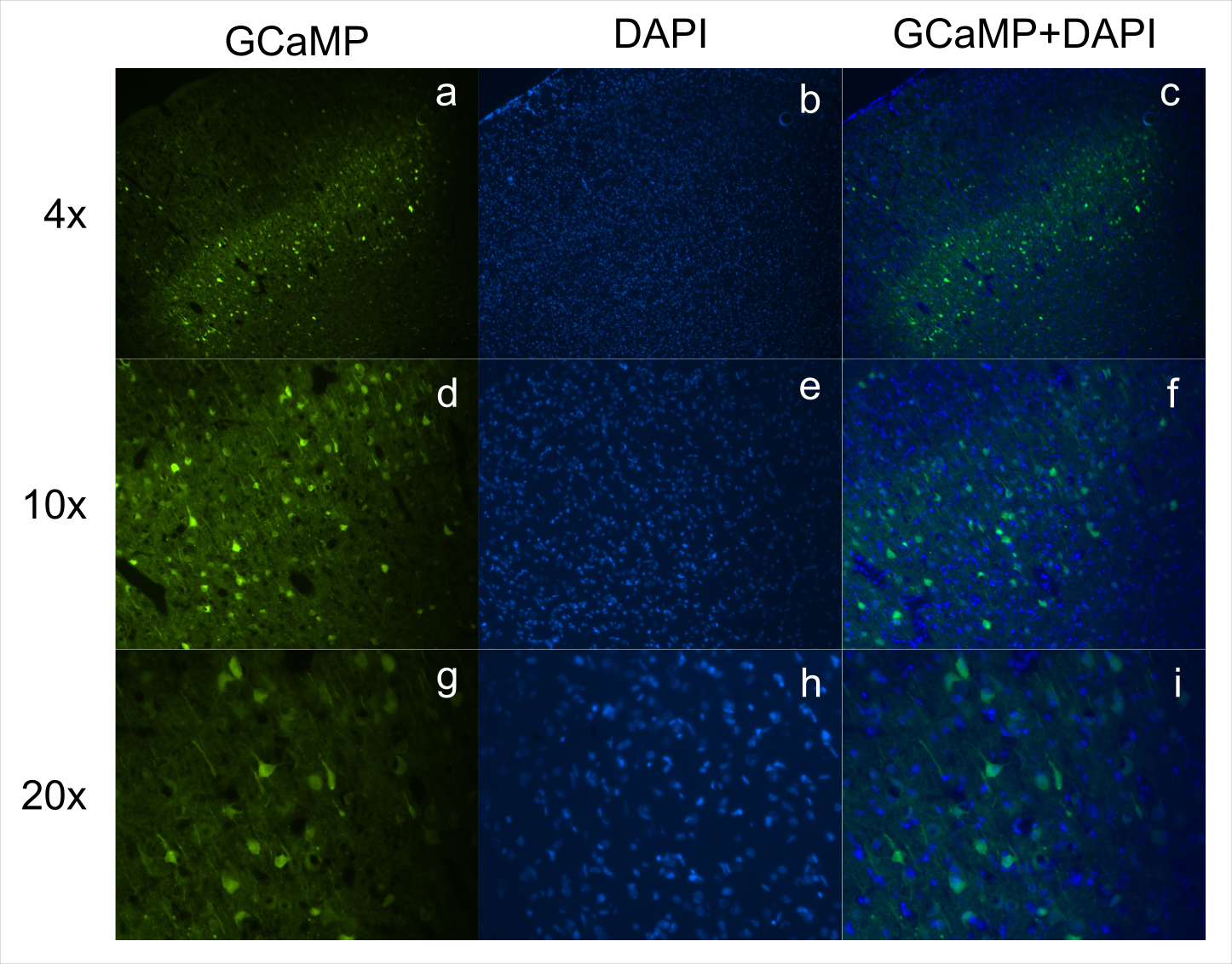
Figure 4
Fluorescence microscopy images from fixed sections of rat brain. Rows represent different magnifications (4x, 10x, 20x). (a,d,g) show expression of GCaMP in green, (b,e,h) show DAPI nuclear staining in blue. Both images are combined in (c,f,i). Cells with GCaMP expression also have a DAPI stained nucleus, indicating cellular integrity.