1668
A novel role of intrinsic astrocytic calcium spikes to mediate brain states through central/dorsal thalamic nuclei1Max Planck Institute for Biological Cybernetics, Tuebingen, Germany, 2Graduate Training Centre of Neuroscience, International Max Planck Research School, University of Tuebingen, Tuebingen, Germany
Synopsis
The astrocytic Ca2+ signal could be simultaneously acquired with either BOLD-fMRI or LFP signal in the rat cortex under anesthesia. Intrinsic astrocytic Ca2+ bursts spikes were detected in the cortex with suppressed spontaneous LFP and negative BOLD fMRI signal. These astrocytic Ca2+ spike events were different from the normal activity-evoked Ca2+ signal and also differentiate themselves from the lesion/stimulation-induced large-scale depolarization or spreading depression given the instantaneous whole-brain pattern and correlation with the thalamic LFP. The intrinsic astrocytic Ca2+ spike may mediate the brain states through the arousal thalamic pathway.
Introduction
Previously, an intrinsic astrocytic calcium spike was detected to be negatively correlated to the BOLD fMRI signal1. By simultaneous calcium with electrophysiology or BOLD fMRI recording, the intrinsic astrocytic Ca2+ spikes coincided with suppressed LFP activity, as well as negative BOLD fMRI signal through the entire cortex. Interestingly, the intrinsic astrocytic Ca2+ spikes could be initiated in bilateral hemispheres at the same time with sensory stimulation. Upon the appearance the intrinsic astrocytic Ca2+ spikes, the whole-brain BOLD fMRI was used to search for the specifically activated brain regions, which may serve as the source to elicit the astrocytic Ca2+ spike. Increased BOLD signal was detected at the central and mediodorsal thalamic nuclei specifically, as well as increased LFP activity upon stimulation. This work indicates that the intrinsic astrocytic Ca2+ signal could play a crucial role to mediate the brain state changes through the arousal thalamic pathways.Methods
All images were acquired with a 14.1 T/26cm horizontal bore magnet (Magnex), interfaced to an AVANCE III console (Bruker) and equipped with a 12 cm gradient set, capable of providing 100 G/cm with a rise time of 150 us (Resonance Research). A transreceiver surface coil was used to acquire fMRI images. Electrodes were placed on the forepaw (FP) to deliver a 1.0 mA pulse sequence (4s, 300μs duration repeated at 3Hz). GCaMP6f 2 was expressed by AAV5 in the FP somatosensory cortex (FP-S1) with Syn or GFAP promoter. Fiber optic (200um) was inserted into the area which expressed GCaMP for calcium-based fluorescent signal recording.Results
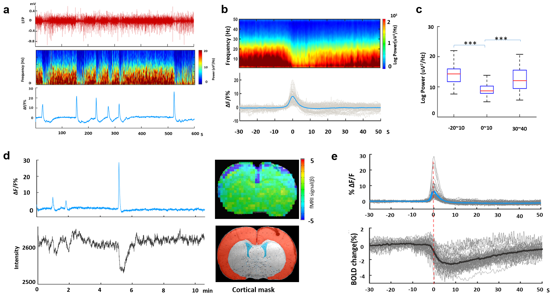
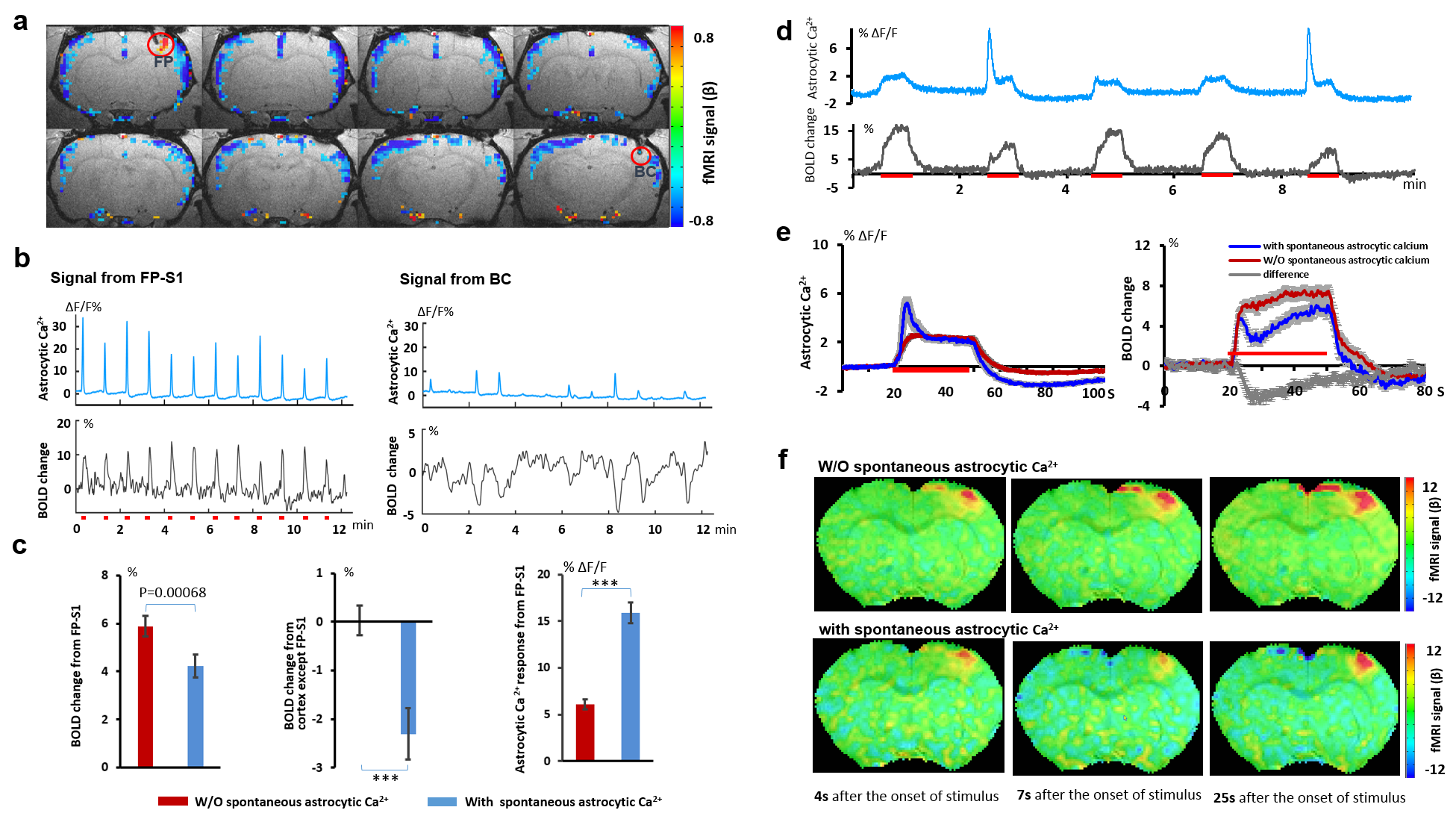
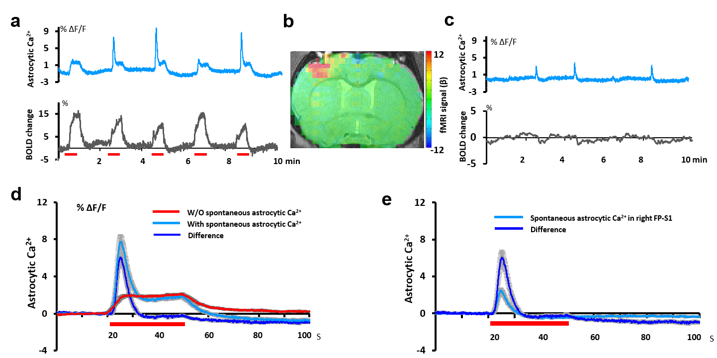
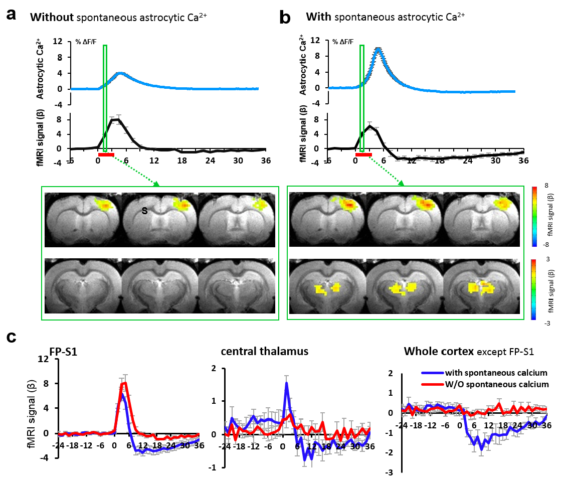
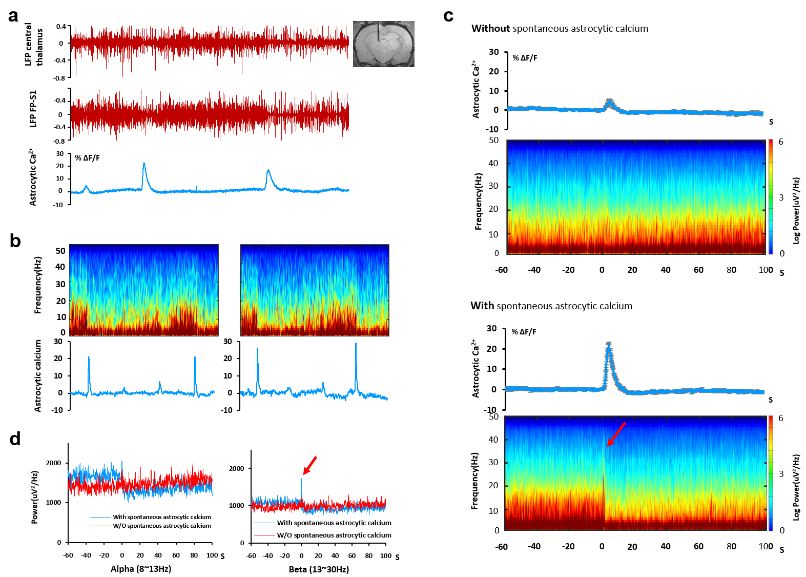
Intrinsic astrocytic Ca2+ spikes were detected in coincidence with the suppressed spontaneous LFP signal, as well as the negative BOLD fMRI signal (Fig 1). This intrinsic astrocytic Ca2+ signal could be initiated by sensory stimulation and was detected from multiple cortical areas, e.g. FP-S1, barrel cortex, or cross hemispheres, with negative BOLD signal through the entire cortex (Fig 2). Upon the 30s electrical unilateral sensory stimulation, the intrinsic astrocytic Ca2+ spikes were detected in a few epochs at both hemispheres, showing spikes with amplitude 2-3 times higher than the normal evoked astrocytic Ca2+ signal (Fig 3). Interestingly, the bilateral intrinsic astrocytic Ca2+ spikes showed no onset delay regardless of the evoked cortical side (Fig 3e), indicating the intrinsic astrocytic Ca2+ spikes are not propagating from the activated cortical areas. The whole-brain BOLD fMRI was simultaneously acquired with the bi-hemispheric astrocytic Ca2+ recording, showing specific thalamic activation upon the appearance of the intrinsic astrocytic Ca2+ spikes but not under the normal evoked astrocytic Ca2+ events (Fig 4). This result indicates that the thalamic activation may directly mediate the large-scale cortical intrinsic astrocytic Ca2+ spike events. Furthermore, simultaneous thalamic LFP recording at the central and mediodorsal region with cortical astrocytic Ca2+ recordings showed that the increased LFP power spectrum density in thalamus upon the intrinsic astrocytic Ca2+ spikes in comparison to the normal evoked astrocytic Ca2+ signal (Fig 5). These results indicates that the intrinsic astrocytic Ca2+ spikes may directly get involved in the brain state changes through the arousal thalamic pathways.Conclusion
The intrinsic astrocytic Ca2+ spikes were detected in the cortex with suppressed spontaneous LFP and negative BOLD fMRI signal. These astrocytic Ca2+ spike events were different from the normal activity-evoked Ca2+ signal and also differentiate themselves from the lesion/stimulation-induced large-scale depolarization or spreading depression given the instantaneous whole-brain pattern and correlation with the thalamic LFP. The intrinsic astrocytic Ca2+ spikes may mediate the brain states through the arousal thalamic pathway.Acknowledgements
This research was supported by the internal funding from Max Planck Society.References
1. Wang et al. ISMRM (abstract ID 2799, 2017). 2. Chen et al. Nature, 499:295-302 (2013).
Figures