1640
Control study of arterial spin labeling and multi-phase enhancement technique in evaluation of renal blood flow in different age populationYimin Wang1, Ailian Liu, Jinghong Liu, Meiyu Sun, Qingwei Song, Ye Li, Anliang Chen, Shifeng Tian, and Hanpei Zhang
1The First Affiliated Hospital of Chengdu Medical College, Chengdu, People's Republic of China
Synopsis
This study is aim to assess ability of arterial spin labeling technique(ASL) and multi-phase enhanced magnetic resonance (MCE-MR) of evaluate renal perfusion in different ages population with normal renal function by 1.5T MRI. Meanwhile, to evaluate difference between ASL and MCE-MR of renal perfusion.
Introduction
ASL(arterial spin labeling) and MCE-MR(Multi-phase contrast enhanced MR)technique are widely recognized as a method which can be quantified by using water protons as the endogenous tracer. ASL can be used in the absolute quantification of RBF(renal blood flow).MCE-MR can assess renal perfusion accroding ER(enhancement ration)Our study aimed to assess relationship between RBF, renal function in population with normal renal function of different ages by 1.5T MRI arterial spin labeling technique(ASL) and MCE-MR. Meanwhile, to evaluate difference between ASL and MCE-MR of renal perfusion.Methods
Retrospectively collected patients who underwent LAVA MCE-MRI and FAIR sequence on 1.5T MR in our hospital during June 2015 to December 2015, and confirmed without renal disease by routine MR images, normal renal function in laboratory examination in a week. The patients enrolled were divided into three groups according to age, group 1 included age between 21-40 years old,group 2 included age between 41-60 years old, group 3 included age between 61-80 years old, then extract 20 cases from each group randomly for our study. RBF were measured and ER were calculated in bilateral kidneys meanwhile(Fig.1-2). eGFR was calculated using the CKD-EPI formula. Gender composition among different group compared by chi-square test.ANOVA test were used to test differences of RBF、ER between all groups. Spearman correlation was used for correlation analysis between age and all parameters, RBF and eGFR,RBF and ER.Pearson correlation was used for correlation analysis between ER and eGFR.Results
①There are significant differences of cortical RBF between 3 groups(P=0.005<0.05),the difference between group 1 and 3 is significantly(P=0.004<0.05).But no significant difference of ER among 3 groups②No correlation between RBF and ER(P=0.911>0.05). ③eGFR of three groups and pairwise comparison have significant differences(P<0.05)④Age showed a negative correlation with RBF,ER and eGFR.⑤No significant correlation between RBF, ER and eGFR.Discussion
Winter[1] considered there is not enough CNR in original images of ASL to distinguish cortex and medulla of kidney,meanwhile,using hydrion as endogenic contrast agent may cause insensitivly in medulla perfusion.Therefore, we chosen cortical RBF compare with early cortical phase ER.There is significant difference crotical RBF between all groups, especially between 21-40years and 61-80years, and there is negative correlation between age and RBF.We suggest that may caused by renal perfusion is decreasing more slowly in the aged.Compared with quantitative RBF values obtained by the ASL, ER is half quantitative indicators merely, the perfusion of information can reflect limited, so there is no certain correlation between RBF and ER. In addition, the contrast agent of MCE-MR is extracellular contrast agent, reflects the exchange of contrast medium and tissue, different from endogenous contrast agent——hydrogen protons in arterial blood of ASL.And in MCE-MR, high-pressure injecting may cause changing of pressure in arteries.Therefore, only ASL can detect diminution in large span age.We also compared our results with others.Differ from us, previous study showed there is correlation between RBF and serum creatinine. Our study chosen CKD-EPI formula for estimating eGFR, give full consideration to the racial and gender.Different TI and tested people may due to inconformity.Conclusion
Either ASL or MCE-MR can reflect renal cortex blood flow reduced with the age growth, but ASL is more sensitive than MCE-MR for changing of renal perfusion caused by aging.Acknowledgements
No acknowledgement found.References
[1]Winter JD, St Lawrence KS, Cheng HL. Quantification of renal perfusion: comparison of arterial spin labeling and dynamic contrast-enhanced MRI. J Magn Reson Imaging, 2011, 34: 608-615Figures
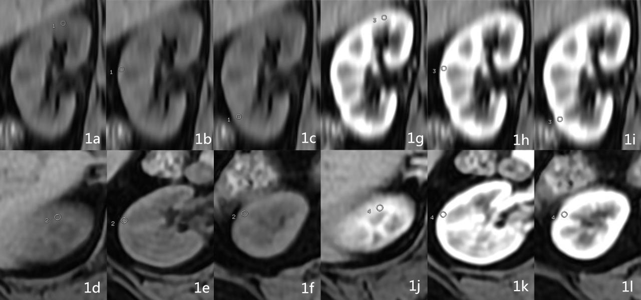
Figure 1a-f: unenchanced phase images. Figure 1g-l: cortical phase images. ROIs were drew in axial images in upper, middle, lower part in each kidney(take right kidney as sample)and corresponding coronal location were record in order to consistent with ROIs in original images of ASL.
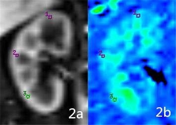
Figure 2a-b: original and pseudo-color images of ASL. ROIs were drew in axial images in upper, middle, lower part in each kidney(take right kidney as sample)refer to coronal MCE-MR images.
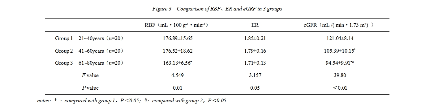
Figure 3 Comparison of RBF、ER and eGRF in 3 groups