1639
Differential Reduction of Repertoire of Functional Patterns in Sensory and Cognitive Neuronal Systems in Propofol Anesthesia1Radiology, Medical College of Wisconsin, Milwaukee, WI, United States, 2Anesthesiology, Medical College of Wisconsin, Milwaukee, WI, United States, 3Biophysics, Medical College of Wisconsin, Milwaukee, WI, United States, 4Neurology, Medical College of Wisconsin, Milwaukee, WI, United States, 5Anesthesiology, University of Michigan, Ann Aobor, MI, United States
Synopsis
We propose that the diversity of distinct functional patterns of the brain can be quantified by the variance explained by the first few principal components of regional voxel functional imaging signals. We report that propofol sedation is associated with a global reduction of repertoire of functional patterns. While sensory-processing-related and high-order cognitive-processing-related brain regions both showed a reduction during propofol sedation, it was the changes in the sensory-processing-related regions that correlated the loss and return of consciousness. The findings provided important insights into anesthetic modulation of different neuronal systems and the neural correlates of consciousness at the systems level.
Introduction
The richness of conscious experience is considered to depend on the formation of dynamic patterns of neuronal coalitions in the brain.1 The diversity, or repertoire, of distinct functional patterns formed within and across neuronal systems reflects the information integration capacity of the brain that is essential in determining the state of consciousness.2 Anesthesia may suppress consciousness by reducing the repertoire of functional patterns in the brain,3 but direct evidence has been scarce. Macroscopic anatomical boundaries have a general, though imperfect, relation to functional boundaries. Here we propose that the diversity of distinct functional patterns of the brain can be quantified by the variance explained by the first few principal components (PCs) of blood-oxygen-level-dependent (BOLD) signals of voxels in each of the well-defined anatomical regions, measured by resting-state functional magnetic resonance imaging (rs-fMRI). The goal of this study is twofold. First, we will test the hypothesis that propofol sedation is associated with a global reduction of repertoire of functional patterns as captured by an overall increase of the variance explained across brain regions. Second, we will determine how the reduction of the repertoire of functional patterns varies in the sensory and high-order cognitive systems of the brain to the changes of consciousness modulated by propofol anesthesia.Methods
rs-fMRI BOLD signals were obtained using a 32-channel head coil from 15 healthy volunteers during four 15-min scans in wakeful baseline, propofol-induced light sedation and deep sedation, and recovery. In light sedation, volunteers showed lethargic response to questions; in deep sedation, volunteers had no response to verbal commands (OAAS score 2-1), indexing unconsciousness. Standard imaging preprocessing procedures were performed. The time series of voxels in each of the 116 anatomical regions, defined by a standard template,4 were analyzed by a principle components analysis. The variance explained by the first five PCs was computed and reported, with an increase in variance explained indicating a reduction in the total number of PCs (thus reduced dynamic functional patterns) and a reduction in variance explained indicating the opposite. Varying the reported number of PCs over a range of values (e.g., 3–12) did not alter the conclusions.Results
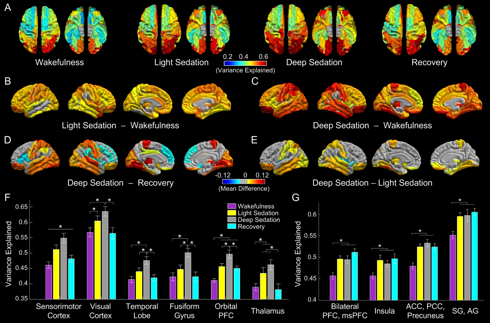
As shown in Fig. 1A, the variance explained by the first five PCs showed a regional inhomogeneity, with relatively small values found in various portions of the anterior brain (e.g., dorsal lateral prefrontal cortex [PFC]) and large values in the posterior regions (e.g., the visual cortex and various parietal regions). There is a trend of increased variance explained with the deepening of sedation, and the trend was moderately reversed in recovery. Compared with wakeful baseline, the group mean difference showed an overall increase of the variance explained in light sedation (Fig. 1B) (no decrease was found), and the increases were further enlarged during deep sedation (Fig. 1C), especially in all the major sensory and motor cortices, the thalamus, the fusiform gyrus, and areas of the orbital PFC. During recovery, prominent reductions in the variance explained showed in the same set of regions, however, with few changes in other sections of the brain and slight increases in the dorsal lateral PFC and insula (Fig. 1D). The transition from light to deep sedation marks the loss of consciousness; this critical change of consciousness was associated with increased variance explained in sensory-processing-related areas but not in high-order cognitive processing areas (Fig. 1E). Region-specific changes in the variance explained, that are consistent with the changes in the state of consciousness, were shown in six sensory-processing-related brain areas (Fig. 1F). Four other brain areas for high-order cognitive processing and multimodal information integration showed an increase in the variance explained in light sedation; however, the same amount of increase sustained during deep sedation and even in recovery, in which consciousness returns (Fig. 1G).Discussion & Conclusion
The extent of ongoing dynamic formation of metastable functional patterns can be represented by variance explained by the first few PCs of regional voxel BOLD fMRI signals. We showed that propofol sedation is associated with a global reduction of repertoire of functional patterns. For the first time, we demonstrated that sensory-processing-related and high-order cognitive-processing-related brain regions exhibit differential changes in the repertoire of functional patterns across the states of consciousness modulated by propofol. While the both sets of regions showed a reduction during propofol sedation, it was the changes in the sensory-processing-related regions that correlated the loss and return of consciousness. The findings of the study provided important insights into anesthetic modulation of neuronal systems of different functional specializations and the neural correlates of consciousness at the systems level.Acknowledgements
Research reported in this publication was supported by grants of the National Institute of General Medical Sciences of the National Institutes of Health under Award Number R01-GM103894 and T32 GM89586. The content is solely the responsibility of the authors and does not necessarily represent the official views of the National Institutes of Health. The authors thank Ms. Lydia Washechek, BA, for editorial assistance.References
1. Werner G. Viewing brain processes as critical state transitions across levels of organization: neural events in cognition and consciousness, and general principles. Biosystems 2009; 96:114–119
2. Tononi G, Koch C.The Neural Correlates of Consciousness. Ann N Y Acad Sci 2008; 1124:239–261.
3. Alkire MT, Hudetz AG, Tononi G. Consciousness and anesthesia. Science 2008; 322:876–880
4. Tzourio-Mazoyer N, et al. Automated anatomical labeling of activations in SPM using a macroscopic anatomical parcellation of the MNI MRI single-subject brain. Neuroimage 2002;15(1):273-89.
Figures