1631
Impact of prospective motion correction in 7T fMRI studies1DZNE, Magdeburg, Germany, 2Institute of cognitive neurology and dementia research (IKND), Magdeburg, Germany, 3Department of Biomedical Magnetic Resonance, Institute of Experimental Physics, Otto-von-Guericke-University, Magdeburg, Germany, 4Center for Behavioral Brain Sciences, Magdeburg, Germany, 5Leibniz Institute for Neurobiology, Magdeburg, Germany
Synopsis
Recently, more fMRI studies are aiming to assess the role of small anatomical regions of the brain in cognitive processes which requires sub-millimiter voxel resolution. At high resolution rigid body motion during the acquisition plays a significant role. The aim of this study is to assess the potential benefits of correcting for subject motion introduced distortions and image degradation prospectively in fMRI studies. This study demonstrates that prospective motion correction increases tSNR and therefore increases sensitivity. These results increment the potential applications of fMRI to unveil, more accurately, the role of smaller parts of the brain in different cognitive processes.
Purpose:
The aim of this study is to assess the potential benefits of correcting for subject motion introduced distortions and image degradation prospectively in fMRI studies. Recently, more fMRI studies are aiming to assess the role of small anatomical regions of the brain in cognitive processes which requires sub-millimiter voxel resolution [1, 2]. At high resolution rigid body motion during the acquisition plays a significant role. We hypothesise that prospective motion correction (PMC) will increase the sensitivity and specificity of the experiments.Methods:
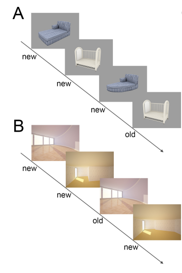
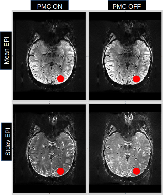
An object-scene mnemonic discrimination task used to investigate functional processing pathways in the medial temporal lobe was used to evaluate the impact of prospective motion correction (Fig 1). To assess the potential benefit of PMC in fMRI studies 7 well trained subjects were scanned (4 females, after written consent); all of them with (PMC ON) and without motion correction (PMC OFF). A mouth piece rigidly connecting a marker to the subject’s upper jaw was built for each subject. The marker - thus subject motion – was tracked by an in-bore camera with 80 frames per second and the current head position and orientation were computed and used to update the imaging volume during acquisition [3-4]. Data was acquired in two separate sessions. The order of the fMRI runs, for each subject, as well as the acquisition of the PMC structural images were counterbalanced to avoid undesired learning effects. In addition two different parallel versions of the functional tasks with matched task difficulty were used. MRI data were acquired using a 7T MR system (Siemens, Erlangen, Germany) with a 32 channel head coil (Nova Medical, Wilmington, USA). The images in PMC ON fMRI sessions were distortion corrected whereas PMC OFF fMRI images were both distortion corrected and realigned using a point spread function mapping method [5]. See table 1 for protocol details. PMC ON and PMC OFF images were processed separately. Images acquired on different days were rigidly co-registered using SPM. Additional preprocessing included slice timing correction and spatial smoothing with a Gaussian kernel (FWHM = 2 mm) [1, 2]. To assess the impact of motion correction in the analysis the temporal SNR (tSNR) in a spherical VOI placed in the left occipital lobe was measured for both PMC ON and PMC OFF images. Measured tSNR values were compared using a standard t-test. Finally, we calculated the number of activated voxels (p<0.001, uncorrected) in both PMC ON and PMC OFF sessions to compare their statistical sensitivities.Results:
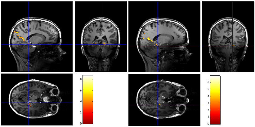
Overall, tSNR values in PMC ON sessions were significantly higher than that in PMC OFF sessions (p<1e-6, Fig 2). This increase in tSNR seems to be driven by the differences between the PMC ON and PMC OFF temporal variations (Fig 3). Furthermore, the statistical sensitivity of detecting difference in the t-contrast of scene versus object is higher in the PMC ON images than in the PMC OFF images: The percentage of significant voxels (p<0.001) increased from 5.95% up to 184% in 6 out of 7 subjects. One example of this increase of significant voxels is shown in Fig 4.Discussion:
Our results illustrate the potential benefit of PMC in functional MRI studies, as demonstrated by a significantly increase in tSNR which in turn increased the statistical sensitivity for subsequent analyses. Our results also suggested that the increase in tSNR in PMC ON images could be due to the absence of a realignment step in PMC ON. During the realignment step, PMC OFF images are co-registered to the first volume; therefore introducing undesired smoothing and volume averaging effects which end up reducing the tSNR and therefore the sensitivity of the measurement. This increase in sensitivity in PMC ON images is consistent with the results reported by Nick et al [6] measured at 3T at lower resolution.Conclusion:
This study demonstrates that PMC increases tSNR and therefore increases sensitivity. These results increase the potential applications of fMRI to unveil, more accurately, the role of smaller parts of the brain in different cognitive processes. In future, a greater number of subjects will be enrolled in the study to increase the statistical power and allow for group comparisons between PMC ON and PMC OFF data.Acknowledgements
This work was supported by the NIH, grant number 1R01-DA021146, and by the Initial Training Network, HiMR, funded by the FP7 Marie Curie Actions of the European Commission, grant number FP7-PEOPLE-2012-ITN-316716.References
1. - Maass A, et al. Nature Communications 5, 5547 (2014)
2. - Berron D, et al. J. Neurosci. 36 (29) 7569-79 (2016)
3.- Stucht D, et al, PloS one. 10 (7) :e0133921 (2015)
4.- Maclaren J, et al, PloS one. 7(11):e48088 (2012)
5. - In, M.-H. & Speck, O. MAGMA. 25, 183–192 (2012)
6. - Todd N, et al, Neuroimage 113, 1-12 (2015)
Figures