1623
Isotropic High Spatial Resolution fMRI Using Accelerated Variable Density Spiral and SLIDER1Center for Biomedical Imaging Research, Department of Biomedical Engineering, School of Medicine, Tsinghua University, Beijing, People's Republic of China, 2Philips Healthcare, Shanghai, People's Republic of China, 3FMRIB Centre, Nuffield Department of Clinical Neurosciences, John Radcliffe Hospital, Oxford, United Kingdom, 4Department of Biomedical Engineering, School of Medicine, Tsinghua University, People's Republic of China
Synopsis
fMRI with high spatial resolution is beneficial for studies in psychology and neuroscience, but is limited by various factors such as prolonged imaging time and low signal-to-noise ratio. In this work, we combined Dual-TRACER method with SLIDER technique to obtain up to 1mm isotropic fMRI image, while maintaining temporal resolution comparable with conventional acquisition. Using the proposed method, finger somatotopy maps were successfully located on a 3T scanner.
Introduction
fMRI with high spatial resolution is beneficial, but is limited by various factors such as prolonged imaging time and low signal-to-noise ratio (SNR). Recently, Dual-TRACER1, based on Temporal Resolution Acceleration with Constrained Evolution Reconstruction (TRACER)2 was proposed. Results show that Dual-TRACER can provide reliable functional images with a high in-plane spatial resolution (1 x 1 mm2) under high acceleration factors. However, the slice thickness in Dual-TRACER is limited due to the SNR and RF profile limitations. Recently, a super resolution based method called SLIce Dithered Enhanced Resolution (SLIDER) was developed. Thinner slice thickness was achieved while maintaining SNR through slice profile shifts in the slice direction3,4.
In this study, the SLDER technique was integrated with Dual-TRACER to improve the spatial resolution in fMRI, achieving up to 1mm isotropic voxels size. The proposed technique was demonstrated in visual stimulus task, as well as finger somatotopy mapping which was only reported at 7T scanner previously5.
Theory
In fMRI, the k-space data of n-th time frame can be formulated in matrix yn=Ψ(xn)=PFSxn [1], where yn is the k-space data from n-th time frame, xn is the image of n-th time frame, S is the coil sensitivity map, F is the Fourier transform and P is the k-space projection onto sampling trajectories. When SLIDER is incorporated, S changes to a generalized encoding map, which contains information of coil sensitivity and RF slice profile encoding. In this work, the SLIDER was incorporated in the Dual-TRACER by changing the encoding function Ψ in equation [1], in order to achieve in-plane and through-slice high resolution.
Methods
In vivo fMRI experiments were conducted with visual stimulus and finger somatotopy mapping tasks. All data were acquired on a Philips 3.0T Achieva TX MRI scanner (Philips Healthcare, Best, The Netherlands) using a 32-channel head coil. Golden angle rotated variable density spiral (VDS) (α=4) was used for acquisition.
Visual Stimulus The visual stimulus paradigm was a block design consisting of 20s of blank screen fixation alternating with 20s of a flashing and rotating checkerboard at 8 Hz. TE=35ms, TR=2s, FOV=200 × 200mm2. Two datasets were acquired: 30 slices with 1 mm3 without SLIDER, and 10 slices of 1 × 1 × 3 mm3 with SLIDER=3 (1mm shift in slice direction). 120 image volumes were acquired in 4 mins with 20-fold undersampling.
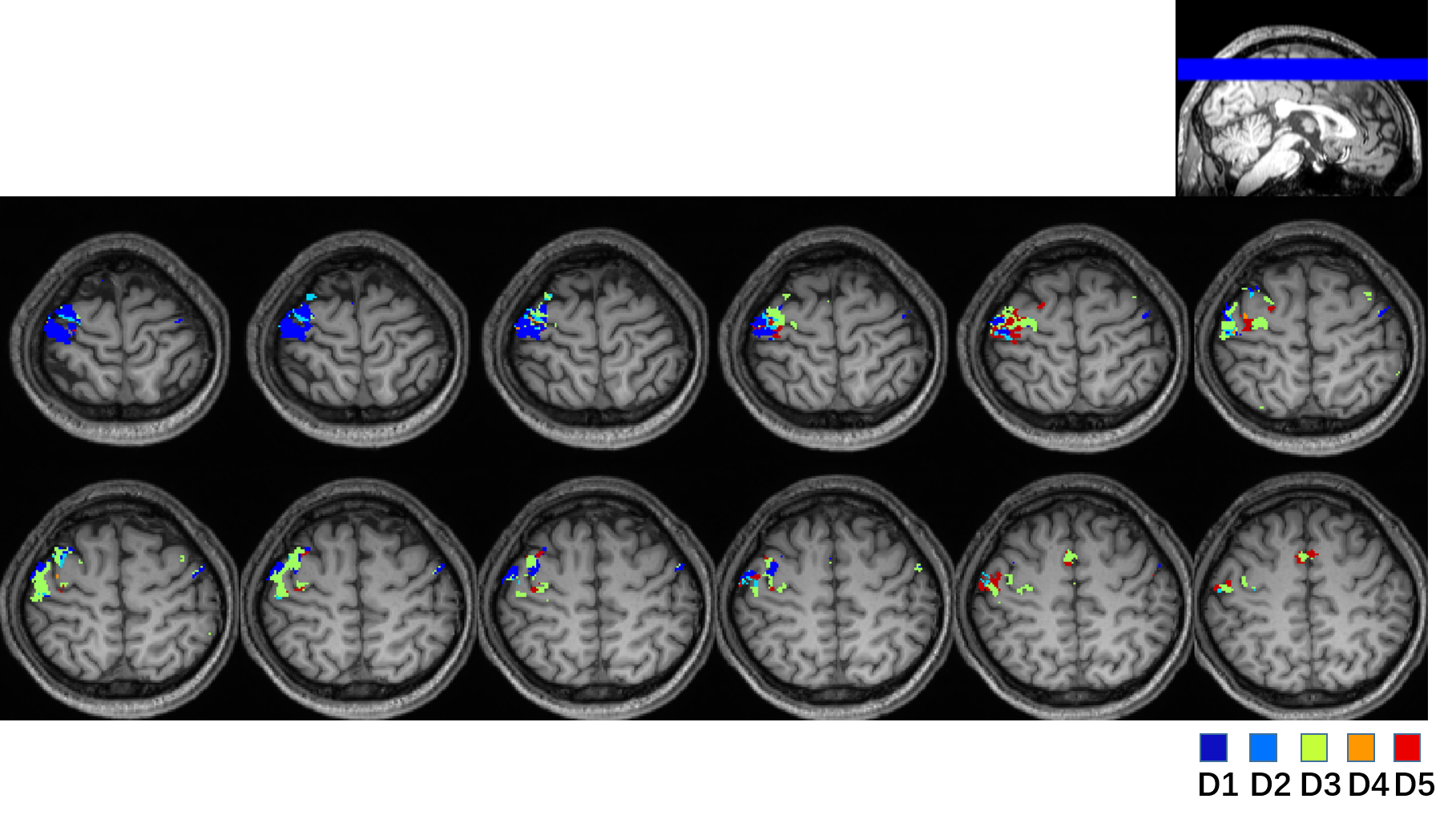
Finger Somatotopy Mapping The stimulus paradigm of the finger mapping task was a block design with 10s rest and 20s finger mapping. Three 10-slice volumes in 1.3 × 1.3 × 3.9 mm3 resolution with a 1.3 mm shift in the slice direction were acquired, achieving a 1.3 mm3 isotropic resolution using SLIDER=3. TE=35ms, TR=2.5s, undersampling factor=12. Five identical sequences for 5 fingers (D1 thumb, D2 index, D3 middle, D4 ring, D5 little) were repeated in a scan time of 20 mins. In each sequence, the two distal phalanges of the subjects' finger were touched by the experimenter5.
Image Reconstruction and Processing All data were reconstructed using Dual-TRACER. For data with shifted volumes, images of isotropic resolution were reconstructed using matrix inversion with Tikhonov regularization. Reconstruction was conducted in MATLAB. Pixel-wise activation maps were calculated using a general linear model (GLM) with familywise error correction (PFWE < 0.05, Z≥5). Functional data were processed using FSL6. FWHM was set to 2mm, as in [5].
Results and Discussion
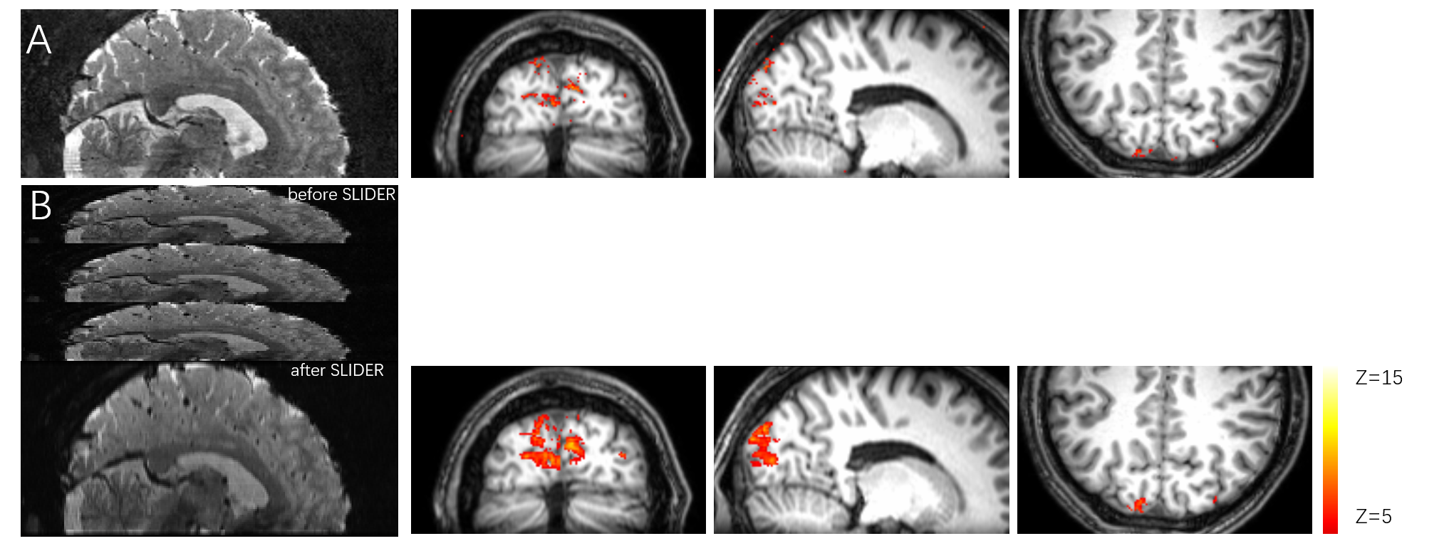
Fig. 1 shows the Dual-TRACER images and activation maps from the visual stimulus experiment without (a) and with (b) SLIDER=3. The SLIDER results can recover the through-plane details compared to the 1 × 1 × 3 mm3 acquisition, and higher SNR can be observed in the images. The activation maps show larger activated area and higher Z score. However, through-slice image blurring can also be observed. Further optimizing of the RF slice profile using SLR pulse and matrix inversion regularization may reduce the image blurring. Fig. 2 shows finger somatotopy mapping results. Through the 1.3mm isotropic resolution, finger activity maps (D1 to D5) can be distinguished from each other on different regions. Due to the long duration and limited SNR, the resolution was limited to 1.3mm isotropic.Conclusion
In this study, a new method combining Dual-TRACER and SLIDER was proposed for high-resolution fMRI with highly undersampling. Results showed that this approach can provide up to 1mm isotropic spatial resolution, The finger somatotopy maps were obtained on a 3T scanner. The improved spatial resolution and maintained temporal resolution are beneficial to clinical research.Acknowledgements
No acknowledgement found.References
[1] Xuesong Li et al. ISMRM 2016. [2] Xu, B et al. Magn Reson Med 2013. [3] Chen, Vu et al, Neoroimage 2015. [4] An T.Vu et.al ISMRM 2016. [5] Martuzzi et.al. Hum Brain Mapp,2014. [6] Smith SM et al. Neoroimage 2004.Figures