1619
Efficacy of double inversion recovery MR imaging in evaluation of the synovium without contrast enhancement1Radiology, Kyung Hee University Hospital at Gangdong, Seoul, Korea, Republic of, 2Radiology, Soonchunhyang University Bucheon Hospital, Buchoen, Korea, Republic of, 3Radiology, Kyung Hee University Medical Center, Seoul, Korea, Republic of
Synopsis
A double-inversion recovery (DIR) sequence simultaneously voids two sources of signals from two different tissues by applying two 180 degree inversion pulses before a signal acquisition. To investigate the efficacy of the DIR image in visualization of the knee synovium without contrast enhancement, contrast-enhanced T1-weighted fat suppressed images (CET1FS) were compared with DIR images in knee MR. Our results showed DIR images were well correlated with CET1FS images.
PURPOSE
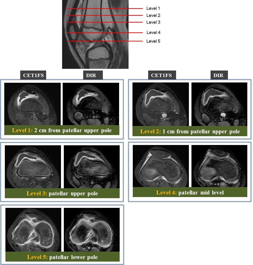
To investigate the efficacy of the double inversion recovery (DIR) image in evaluation of the synovium at the knee joint without contrast enhancement.METHODS
Thirty-three knees from 32 patients (mean age, 41.35 years) were included. After obtaining axial DIR and contrast-enhanced T1-weighted fat-saturated (CET1FS) images, two radiologists independently evaluated their agreements (consistency) in visualization and distribution of the synovium on DIR and CET1FS at each five level of the knee joint using a four-point visual scaling system, and also determined a location with the thickest synovium. The maximal synovial thickness on each sequence was measured by consensus. The synovium-to-effusion signal ratio (SER) and the synovium-to-bone signal ratio (SBR) were assessed at each level. Paired t-tests were performed for comparison of DIR and CET1FS on the maximum synovial thickness, SER, and SBR.RESULTS
Inter-observer agreement was good (κ = 0.736) for the four-point scale and was very good for the location of the thickest synovium on DIR and CET1FS (κ = 0.955 and 0.954, respectively). Inter-sequential agreements for the location of the thickest synovium were very good (R1, κ = 0.845; R2, κ = 0.828). The synovial thickness on each sequence showed a statistically excellent correlation (r=0.872). Mean SERs and mean SBRs were significantly higher at DIR than CET1FS (p< 0.05).CONCLUSION
The DIR image for evaluating the synovium at the knee joint showed good correlation with the CET1FS image. Therefore, DIR may be a one of useful MR techniques for evaluating the synovium of the knee without contrast enhancement.Acknowledgements
This study has received funding by the Basic Science Research Program through the National Research Foundation of Korea (NRF), funded by the Ministry of Education, Science and Technology (2009-0089314).References
1, Jahng GH, et al. Med Phys. 2011;38(5):2579-2585.
2. Kim JH, et al. J Magn Reson Imaging. 2014;39(1):51-58.
3. Krasnokutsky S, et al. Arthritis Rheum. 2011;63(10):2983-2991.
4. Ostergaard M, et al. Arthritis Rheum. 1997;40(10):1856-1867.
5. Loeuille D, et al. Osteoarthritis Cartilage 2011;19(12):1433–1439.
Figures