1616
Age estimation of soft tissue hematomas using 3.0 T MRI: A feasible approach1Institute of Forensic Medicine, Medical University of Graz, Graz, Austria, 2Ludwig Boltzmann Institute Clinical Forensic Imaging, Graz, Austria, 3Division of Neuroradiology, Vascular and Interventional Radiology, Department of Radiology, Medical University of Graz, Graz, Austria, 4Institute of Medical Engineering, Graz University of Technology, Graz, Austria, 5Institute of Forensic Medicine, University of Basel - Health Department Basel, Basel, Switzerland
Synopsis
Forensic
imaging requires reliable analysis of hematomas and their resolution as they
can have significant medico-legal consequences. In 30 healthy volunteers
autologous blood was injected into the subcutaneous fatty tissue of the thigh
and repetitive MRI scans of the artificial hematoma were performed on a 3T
scanner. Due to the contrast behavior three relevant age ranges were defined
and validated. The generated approach correctly classified all tested hematomas
with an age less or equal 24 hours. The development of a reliable and objective
MR-based age estimation method of hematomas is in the interest of justice.
Purpose
As subcutaneous hematomas are usually not relevant for clinicians only limited knowledge and experience exists regarding the detection and dating of traumatic lesions in subcutaneous fatty tissue using MRI. Dating soft tissue injuries such as hematomas is particularly important for the forensic reconstruction of criminal acts, e.g. child abuse cases, where accurate timing of injuries can include or exclude potential suspects.1,2 However, visual assessment of hematoma color, the currently used method for estimating hematoma age, is unreliable due to its great variability2, and is additionally affected by individually varying color perception.3 Consequently, dating of hematomas is difficult, due to a lack of an objective method. First MRI studies showed that the contrast behavior of hematomas could be used to get objective information on hematoma characteristics and that the degeneration of hematomas follows a mono-exponential curve.4,5 The aim of this study was to generate and validate a first feasible approach for hematoma dating using MR contrast changes between blood and subcutaneous fatty tissue over time in order to reliably estimate their age.Methods
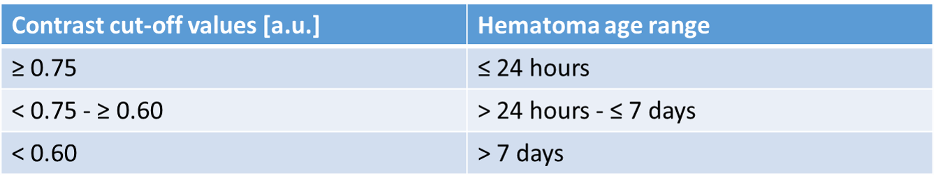
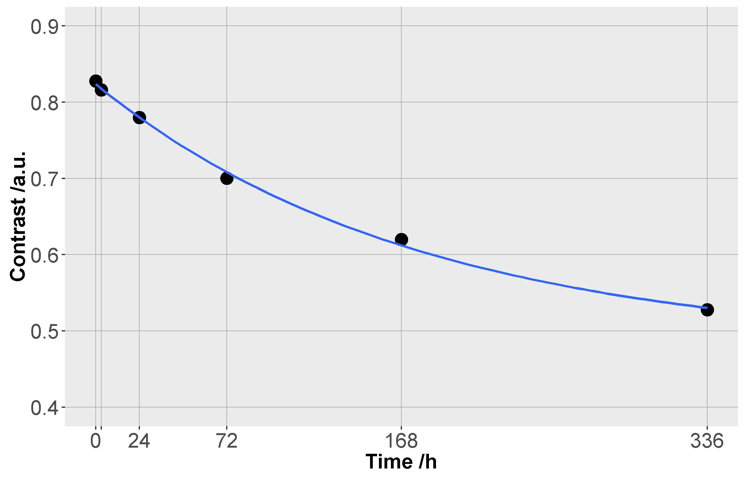
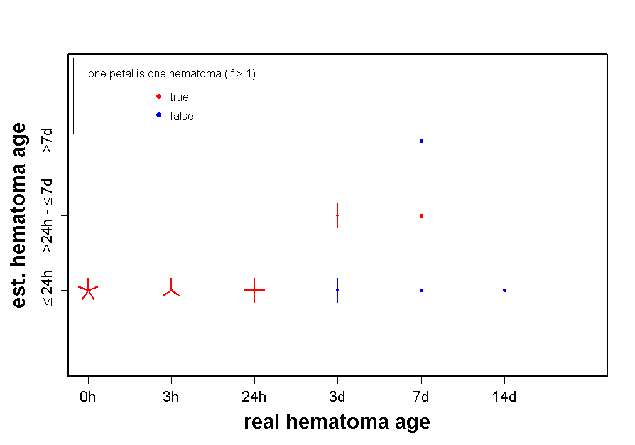
In a study group of 30 healthy volunteers (18m, 12f, age: 26.3 ± 3.8y) without coagulation disorders or medication influencing blood clotting, 4 mL of autologous blood were injected into the subcutaneous fatty tissue of the thigh after a basis MR-scan. The artificial hematoma was scanned repetitively at different points in time (directly after the injection and 3h, 24h, 3d, 7d and 14d later). All measurements were performed on a 3T scanner (TimTrio, Siemens) using a CPC-multifunctional coil (Noras MRIproducts). The MR sequence protocol consisted of a PDw TSE SPAIR (TR/TE 3400/11ms, resolution 0.5x0.5x1.5mm³), a TIR (5xTI) and a MSE (16 echoes) in axial and oblique orientation. Data evaluation was done using a DICOM viewer software (OsiriX 4.1) by measuring signal intensities in 9 ROIs (0.4cm²), three each in the hematoma, fat and muscle tissue. After pooling the measurements of each tissue the contrast coefficient6 was calculated for the different points in time. For further evaluation contrast coefficients were averaged at the single points in time and approximated by a mono-exponential fit according to following equation4: y = a * e-x/t + c. Based on the fitting curve three contrast cut-off values for forensic relevant age ranges were defined (Tab.1). The suitability of this approach was validated with a test group of another 20 artificial hematomas of different age which were created and evaluated as described above. The hematoma age was blinded for the evaluator. Finally, the match of the real and the estimated hematoma age was assessed.Results
Fig.1 shows an example of an artificial hematoma in a PDw sequence at different points in time. In general, the higher the contrast between hematoma and fatty tissue, the more recent was the time of origin of the hematoma. The analysis of MR contrast changes over a time period of two weeks showed an exponential decay (Fig.2). According to the generated age estimation approach the hematoma age of the test hematomas were correctly assigned to the corresponding age range in 75% of the cases. All hematomas with an age less or equal 24 hours were correctly classified by the presented approach (Fig.3).Discussion
Artificial hematomas investigated over two weeks showed differences in the contrast behavior depending on their time of origin. Considering these observations an age estimation approach for hematomas was build and validated using a test group. The majority of the tested hematomas was correctly classified, but most notably was the accurate age estimation of all hematomas with an age ≤ 24 hours. In forensically relevant cases especially the detection of recent bruises is important as it is a well-known fact, that not every hematoma is immediately visible. Therefore, the exact detectability and age estimation of hematomas can have significant medico-legal consequences.Conclusion
The applicability and improvement of this generated approach will be further investigated including additional factors (e.g. other sequences, a higher number of hematomas of different ages), although already these results suggest that it is a suitable and objective method to dependably assess the age of bruises particularly in cases of living victims. Nevertheless, one has to take into consideration that real hematomas might differ concerning their degeneration process. Therefore, in a subsequent study with real hematomas the generated approach has to be validated. In the future, an objective MR-based age estimation method of hematomas could improve the forensic expert’s report in court and may ensure a higher degree of legal certainty.Acknowledgements
No acknowledgement found.References
1. Langlois NEI. The science behind the quest to determine the age of bruises – a review of the English language literature. Forensic Sci Med Pathol. 2007;3(4):241–251.
2. Pilling ML, Vanezis P, Perrett D et al. Visual assessment of the timing of bruising by forensic experts. J Forensic Leg Med. 2010;17(3):143-149.
3. Hughes VK, Ellis PS, Langlois NEI The perception of yellow in bruises. J Clin Forensic Med. 2004;11(5):257-259.
4. Neumayer B, Hassler E, Petrovic A et al. Age determination of soft tissue hematomas. NMR Biomed. 2014;27:1397–1402.
5. Hassler EM, Ogris K, Petrovic A et al. Int J Legal Med. 2015;129:317–324.
6. McRobbie DW, Moore EA, Graves MJ et al. MRI from Picture to Proton. Cambridge University Press, 2007.
Figures