1608
Cartesian UTE with echo time below 0.5 millisecond1Institute for Biomedical Engineering, ETH, Zurich, Switzerland
Synopsis
MRI with sub-millisecond TE typically uses non-Cartesian sampling strategies such as radial UTE leading to increased sensitivity to eddy currents and a high computational burden. We report that TE values in the range so far reserved for radial UTE can be reached with Cartesian sampling using a gradient coil dedicated for muscoskeletal applications in humans.
Introduction
Sub-millisecond echo times (TE) are necessary in many medical MRI applications targeting bones, cartilage, tendons and ligaments, as well as short-T2 components in white matter such as the myelin water. The minimum TE in conventional MRI methods, which sample the k-space on a Cartesian grid, is related to the minimum duration of the phase encoding gradient pulse. With typical gradient systems and the resolution of 0.5 mm, which is standard in muscoskeletal MRI, TE of Cartesian sequences is situated well above one millisecond and could be squeezed down to about 0.8 ms only for the central k-space part by careful sequence optimization (1). Due to this limitation, MRI with sub-millisecond TE almost exclusively uses non-Cartesian sampling strategies such as radial UTE (2). This has the drawback of a less efficient sampling, increased sensitivity to gradient delays and eddy currents and presents a higher reconstruction burden, especially when combined with sensitivity encoding. Here we report that TE values in the range so far reserved for radial UTE can be reached with Cartesian sampling using a gradient coil dedicated for muscoskeletal applications in humans.Methods
A custom-built gradient coil of 33 cm inner diameter has been inserted into the whole body gradient coil of a 3T scanner (Achieva, Philps, The Netherlands). The coil is connected to Copley 787 amplifiers which are normally used for the standard whole body gradient configuration of this system. A maximum gradient of 100 mT/m can be reached with a slew rate of 1200T/m/sec. A home-built transmit-receive quadrature birdcage coil of 28 cm inner diameter, dedicated for head imaging, was used for this study. The sequence was the manufacturer’s standard implementation of 3D gradient echo imaging with non-selective RF pulses and partial echo sampling (10% of the k-space on the “left hand” side). Using a 30-microsecond block RF pulse of 7° flip angle and TR of 3 ms, the echo time of 0.5 ms could be used with 0.5 mm isotropic resolution. To maintain a sufficient signal-to-noise ratio despite a suboptimal filling factor for knee scanning with the present coil, the in-vivo experiment was carried out with a limited resolution of 1x1x2 mm and TE=0.45 ms. The actual response of the gradients has been measured by a field camera (Skope, Switzerland).Results and discussion
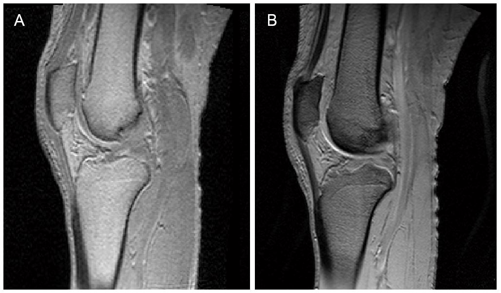
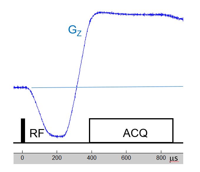
Images of a knee of a healthy male adult are presented in Fig. 1. The image acquired with TE=0.45ms contains strong signal from all types of tissues except for the cortical bone which remains close to the noise level. In particular, the quadriceps tendon and the patellar ligament visible in the central sagittal plane (Fig 1A) have intensity equal to that of the muscle, which is in contrast to the gradient echo at 4.6ms (Fig 1.B) where these structures are dark. The images are not affected by aliasing of out-of-FOV parts of the leg due to frequency encoding of the longitudinal direction. With radial scanning anti-aliasing would require an increased number of projections and a longer scan time. It remains to be investigated if the slight enhancement of contours orthogonal to the z-axis (direction of the readout gradient) is caused by the slight overshoot of the z gradient, which is visible in the field camera measurement (Fig.2), and whether this can be corrected by 1-dimensional k-space gridding. However, a simple delay of the gradient response is certainly less harmful than in radial UTE, as it only leads to a linear phase ramp across the image, an effect automatically removed by the reconstruction.Conclusion
Images with echo time below 0.5 milliseconds having contrast properties similar to radial UTE can be acquired with a standard Cartesian partial gradient echo sequence using a dedicated gradient insert providing 100mT/m with a slew rate of 1200T/m/s. The benefit of this approach compared to radial UTE is a higher sampling efficiency, reduced sensitivity to gradient response imperfections, shorter minimum scan time, avoidance of longitudinal aliasing and the simplified use of sensitivity encoding.Acknowledgements
No acknowledgement found.References
1. Deligianni, X., et al. "High-resolution Fourier-encoded sub-millisecond echo time musculoskeletal imaging at 3 Tesla and 7 Tesla." Magnetic resonance in medicine 70.5 (2013): 1434-1439.
2. Robson, Matthew D., et al. "Magnetic resonance: an introduction to ultrashort TE (UTE) imaging." Journal of computer assisted tomography 27.6 (2003): 825-846.
Figures