1606
Fat Fraction Histogram Analysis: Insights into the Biology of Lipomatous Tumors1Department of Radiation Sciences, Umeå University, Umeå, Sweden, 2Department of Radiology, Uppsala, Sweden, 3Department of Neuroradiology, Karolinska University Hospital, Stockholm, Sweden, 4Department of Oncology and Pathology, Karolinska Institute and Karolinska University Hospital, Solna, Sweden, 5Department of Orthopaedic Surgery, Karolinska University Hospital, Solna, Sweden, 6Department of Medical Radiation Physics, Karolinska University Hospital, Stockholm, Sweden, 7Department of Clinical Science, Intervention and Technology, Karolinska Institute, Stockholm, Sweden
Synopsis
Fat content evaluation in soft-tissue tumors is based on visual grading of T1-weighted images with uncertain accuracy and inter-rater variability. We used a multi-point Dixon technique to quantitatively measure the distribution of proton density fat fraction in different lipomatous tumors. The effect of radiotherapy on fat content in a myxoid liposarcoma was also assessed, with histograms revealing a distinct alteration. Our data give supporting evidence that maturation of tumor cells is the cause for the lipoma-like areas seen after radiotherapy of myxoid liposarcomas. On a voxel level (2×2×2 mm3) various lipomatous tumors exhibited different fat fraction histograms.
TARGET AUDIENCE
Clinicians interested in soft-tissue tumors, radiotherapy and Dixon techniques.PURPOSE
Soft-tissue sarcomas are malignant tumors moderately sensitive to radiotherapy, with the exception of the highly radiosensitive myxoid liposarcomas. Using subjective visual grading on T1-weighted images these lipomatous tumors contain approximately 0-25% fat1. Interestingly, following radiotherapy the total fat amount often increases, while the tumor volume decreases. Two hypotheses have been proposed; either the more mature fat cells are left unaffected or radiation induces a maturation of tumor cells2. Since existing MRI studies assessing fat content in soft-tissue tumors are based on visual grading the accuracy is uncertain and there is inter-rater variability1,3. Albeit subjective, such grading is somewhat helpful in differentiating lipomatous tumors, e.g. liposarcomas (malignant), atypical lipomas/well-differentiated liposarcomas (ALT/WD; semi-malignant) and lipomas (benign)3. In this study we used a multi-point Dixon technique to quantitatively measure the distribution of proton density fat fraction (FF; i.e. fat/fat+water ratio per voxel) in different lipomatous tumors.METHODS
The study was approved by the local ethics committee and informed consent was given by all subjects. A Siemens Verio 3T system and an 18-channel receive body coil was used to acquire 3D spoiled gradient multi-echo images with ten echoes. Relevant scan parameters were TE1/ΔTE/TR=1.88/1.84/21 ms, FA=3° (to minimize T1 bias), matrix=128, FOV=256 mm (rectangular FOV and number of slices was varied based on anatomy and tumor extent), voxel 2×2×2 mm3, pixel BW=1500Hz and NSA=1. A multi-scale graph-cut algorithm was used for fat-water separation4. The MRI sequence was validated on fat-water phantoms with known fat fractions. T1-weighted and T2-weighted images were also included in the protocol. Four patients were examined (one lipoma, one ALT/WD liposarcoma and two myxoid liposarcomas). One myxoid liposarcoma (case #1) was examined pre- and post-radiotherapy. A radiologist delineated each tumor and fat fraction was measured for the total tumor volume. In the lipoma patient, subcutaneous tissue was also delineated at the level of the tumor and measured for reference. All tumors were confirmed by histopathology. Prior to radiotherapy, fine-needle aspiration cytology was also performed in the myxoid liposarcoma.RESULTS
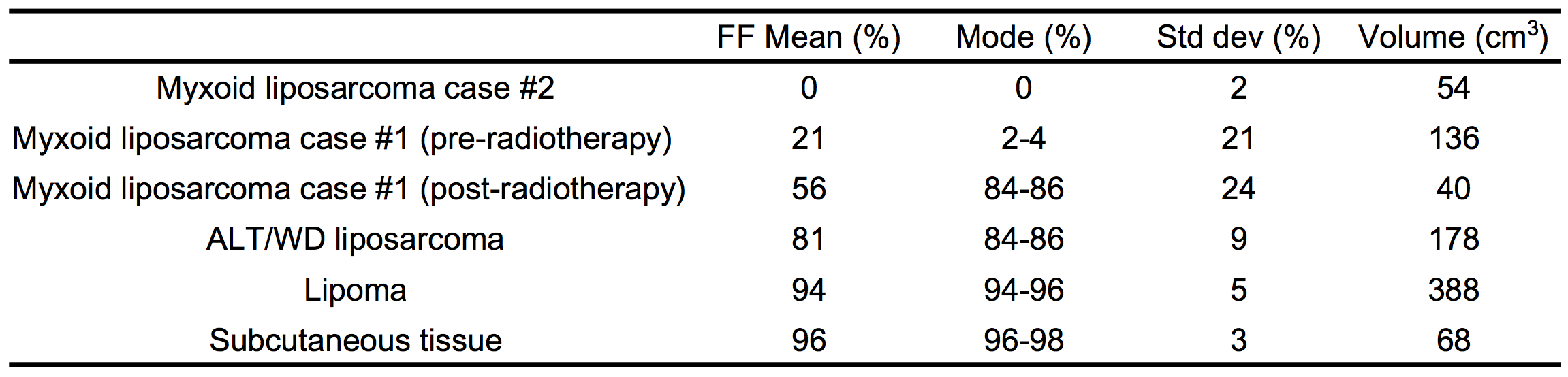
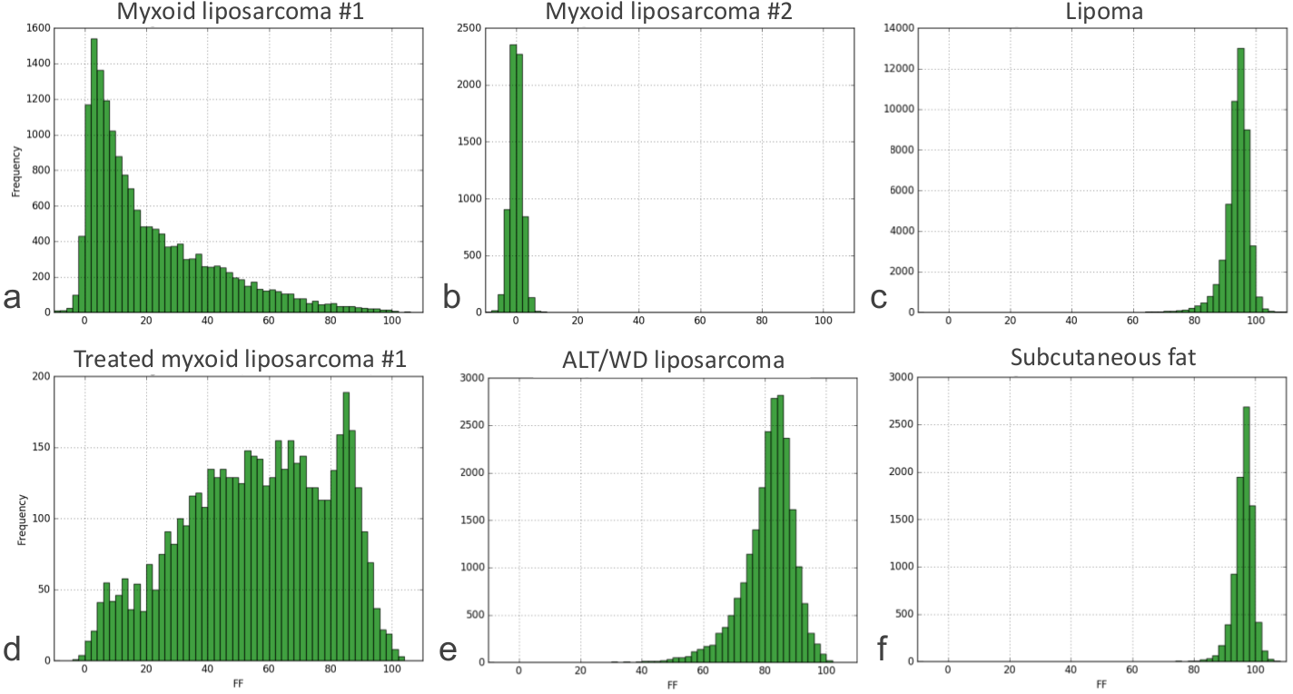
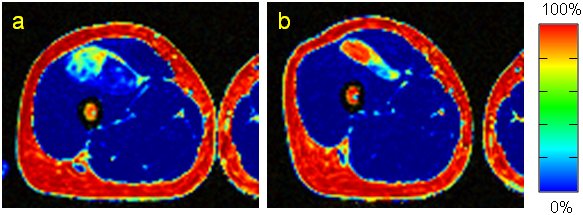
Mean, mode, and standard deviation of intra-tumor FF distributions and tumor volumes are presented in Table 1, and histograms in Fig. 1. Axial FF images, pre- and post-radiotherapy of the myxoid liposarcoma, are presented in Fig. 2. Following radiotherapy the histogram clearly changed in the myxoid liposarcoma, and the histograms also differed between the various lipomatous tumors.DISCUSSION
By using a multi-point Dixon technique to objectively quantify fat fraction in lipomatous tumors, histogram analysis provided new information on these tumors. Following radiotherapy of a myxoid liposarcoma the tumor volume decreased, while both mean FF and mode clearly increased. Most important, voxels containing a much higher fraction of fat appeared, i.e. a distinct transformation of the histogram (Fig. 1a,d). This strongly supports that maturation of tumor cells is the cause for induced lipoma-like areas seen on histopathology2. In addition, following radiotherapy few voxels had a very high FF (>90%), which instead was seen in the lipoma and in subcutaneous tissue (Fig. 1e,f). This would be in keeping with post-radiated induced fat not being regular benign fat, since the typical translocation involving t(12;16) found in myxoid liposarcomas still is evident in induced mature fat5. The pre-radiated myxoid liposarcoma had few voxels with FF >50%, which on contrary was seen in the ALT/WD liposarcoma and lipoma. Both these tumors had a much higher mean FF, but between them there was still a difference in mean, mode and standard deviation (Fig. 1c,f). This could reflect the different tumor biology, which is supported by that the genetic marker MDM2 often distinguishes ALT/WD liposarcoma from lipoma6. Further studies are needed to compare other types of lipomas (containing fat necrosis, chondroid metaplasia etc.) with ALT/WD liposarcoma. In concordance with the literature the myxoid areas were sensitive to radiotherapy, seen as a highly diminished number of voxels with very low FF <10% (Fig. 1d). Occasionally a round cell component can be seen in myxoid liposarcomas. According to Löwenthal et al this component is correlated to intermediate signaling areas on T2-weighted images, which was not seen in the myxoid liposarcomas in this study7. It would be of interest to investigate if myxoid liposarcomas with round cell components have a different histogram appearance, since the round cell component is an important prognostic factor for local recurrence and metastasis8.CONCLUSION
Our data give further supporting evidence that maturation of tumor cells is the cause for the lipoma-like areas seen after radiotherapy of myxoid liposarcomas. On a voxel level (2×2×2 mm3) various lipomatous tumors exhibited different fat fraction histograms.Acknowledgements
No acknowledgement found.References
1. Wortman JR, Tirumani SH, Tirumani H, et al. Neoadjuvant radiation in primary extremity liposarcoma: correlation of MRI features with histopathology. Eur Radiol. 2016;26(5):1226-1234
2. Engstrom K, Bergh P, Cederlund CG, et al. Irradiation of myxoid/round cell liposarcoma induces volume reduction and lipoma-like morphology. Acta Oncol. 2007;46(6):838-845
3. Einarsdottir H, Soderlund V, Larsson O, Mandahl N, Bauer HC. 110 subfascial lipomatous tumors. MR and CT findings versus histopathological diagnosis and cytogenetic analysis. Acta Radiol. 1999;40(6):603-609
4. Berglund J, Skorpil M. Multi-scale graph-cut algorithm for efficient water-fat separation. Magn Reson Med. 2016, in press [doi: 10.1002/mrm.26479]
5. Wang WL, Katz D, Araujo DM, et al. Extensive adipocytic maturation can be seen in myxoid liposarcomas treated with neoadjuvant doxorubicin and ifosfamide and pre-operative radiation therapy. Clin Sarcoma Res. 2012;2(1):25
6. Clay MR, Martinez AP, Weiss SW, Edgar MA. MDM2 and CDK4 Immunohistochemistry: Should It Be Used in Problematic Differentiated Lipomatous Tumors?: A New Perspective. Am J Surg Pathol. 2016
7. Lowenthal D, Zeile M, Niederhagen M, et al. Differentiation of myxoid liposarcoma by magnetic resonance imaging: a histopathologic correlation. Acta Radiol. 2014;55(8):952-960
8. Lemeur M, Mattei JC, Souteyrand P, Chagnaud C, Curvale G, Rochwerger A. Prognostic factors for the recurrence of myxoid liposarcoma: 20 cases with up to 8 years follow-up. Orthop Traumatol Surg Res. 2015;101(1):103-107
Figures