1594
Age estimation using MR imaging of the third molar teeth and the medial clavicular epiphysis: Validation of a multifactorial approach1Ludwig Boltzmann Institute Clinical Forensic Imaging, Graz, Austria, 2BioTechMed, Graz, Austria, 3University Center of Legal Medicine Lausanne-Geneva, University Hospital of Lausanne, Switzerland, 4Institute of Medical Engineering, Graz University of Technology, Austria, 5Institute of Forensic Medicine, Medical University of Graz, Austria, 6Institute for Computer Graphics and Vision, Graz University of Technology, Austria, 7Institute of Forensic Medicine, University of Basel - Health Department Basel, Switzerland
Synopsis
High migration rates in the last years put forensic age estimation of living people at the forefront of forensic research. The established standard for age estimation uses images acquired with ionizing radiation, therefore radiation-free alternatives such as MRI are currently of high interest. In this study a CT based multifactorial approach that combines wisdom teeth and clavicles was validated with MRI data. The sensitivity to estimate subjects under 18 years of age as minors with MRI lies above 93%. Our results showed that MR could replace the CT based multifactorial Approach.
Purpose
The demand for forensic age estimations in living persons increased in the last years due to the large number of people migrating from countries with no reliable documentation of the birth date. To date age estimation relies on the evaluation of the developmental stages of the teeth and the bones using radiological methods based on ionizing radiation. As the use of ionizing radiation without medical indication is ethically controversial and not permitted in many countries, the search for radiation- free alternatives is in the focus of forensic research. Recently Bassed et al.1 published a multifactorial approach using CT images of the wisdom teeth and the medial clavicular epiphysis (cohort age range: 15-25 years). Their purpose was to provide a model with narrower confidence intervals for the estimated age range than when the single regions were used. They provided linear regression formulas to calculate a range with a lower and upper limit for the age determination. The aim of this study was to validate this approach using MR imaging.Materials & Methods
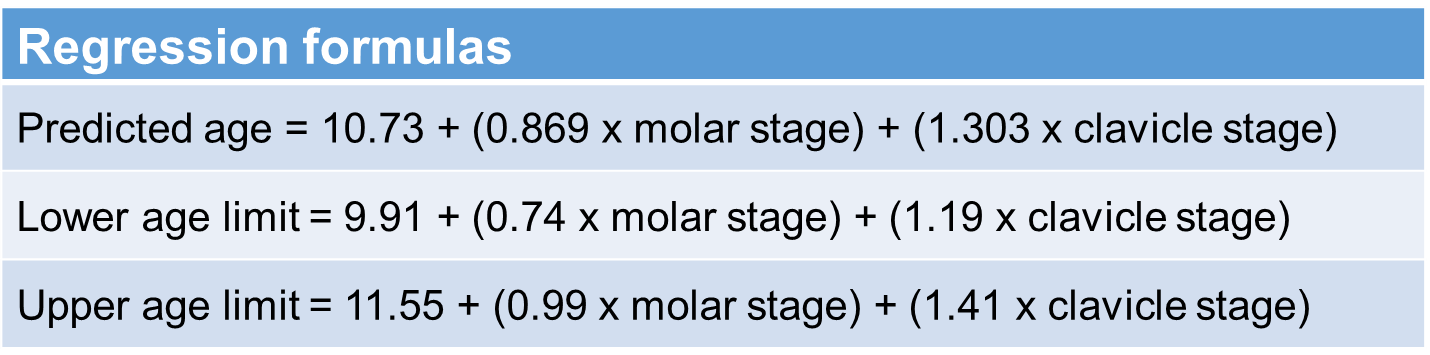
150 male subjects (age 13.01-24.98y, mean 19.9±3.4y) were investigated for this study. MRI of the clavicles and the teeth using a head/neck coil for the clavicles and a multipurpose coil (Noras) for the teeth was performed at 3T (TimTrio, Siemens). The following sequences were applied: a) clavicles: 2D T2wTSE (TR/TE 2905/65ms, 1x1x2mm³), 3D T1wVIBE FS (TR/TE 9.77/3.72ms, 0.9x0.9x0.9mm³) and b) teeth: 3D PDwTSE FS (TR/TE 172/10ms, 0.6x0.6x1.0mm³), 3D T2wCISS (TR/TE 5.41/2.33ms, 0.6x0.6x1.0mm³). Evaluation of the lower third molars and the clavicles were performed by a dentist and a radiologist, respectively, according to defined stages2,3. Calculation of the lower and the upper limit and the predicted age according to the regression formulas provided by Bassed et al.1 was done. (Tab.1) The validation of this approach was investigated using descriptive statistics and calculation of sensitivity and specificity for determining the legally important age limit of 18 years. Additionally, the difference of the mean estimated age of the single regions based on available reference values4,5 and the difference of the predicted values of the multifactorial approach have been compared to the chronological age.
Results
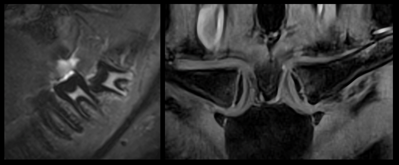
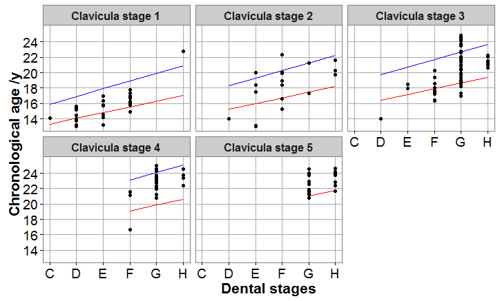
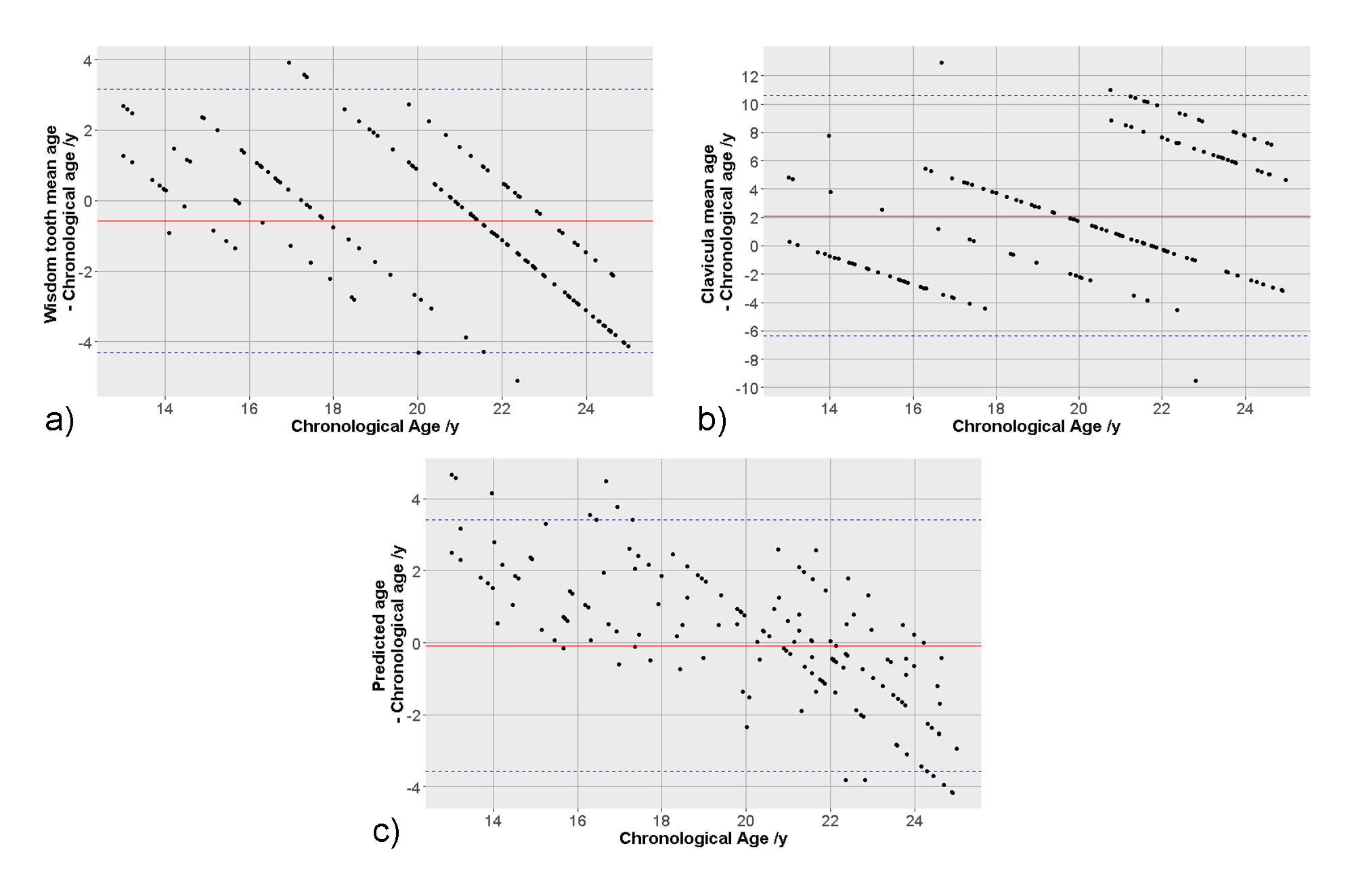
Fig.1 shows representative MR images of the two body regions. The chronological age of 66% of the subjects was within the calculated multifactorial age range (Fig.2). 20.7% were under the lower age limit and 13.3% were above the upper limit. Mainly subjects aged 13 and 14y lay below the limits (16 of 31). Excluding these subjects raised the overall rate to 73.5%. All subjects who were above the upper limit were adults over 18y with a calculated lower age limit above 18y. Concerning the age range regarding the legally important age limit of 18y, 44 of 47 subjects with a chronological age <18y had a calculated lower age limit < 18y corresponding to a sensitivity to estimate a minor subject to be <18y of 93.6%. The specificity was 86.4%, i.e. 86 of 103 adults were correctly classified as adults. The comparison of the chronological age with the predicted age showed a mean difference of 0.08y with a standard deviation of ±1.78y. The single regions displayed a mean difference of 0.58y (±1.91y) and 2.11y (±4.32y). (Fig.3)Discussion
The evaluation showed that only two thirds of the subjects were within the calculated range. Especially subjects under 15 years were below the lower range limit. This could be explained by the fact that Bassed et al.1 used a cohort from 15 to 25 years. However, the legally important question is if the estimated lower limit for minors lies under 18 years. Regarding this question the approach showed good results with a high sensitivity. Also specificity which indicates if an adult is correctly estimated as adult, showed a reasonable result, particularly when considering that an underestimation is not a disadvantage for examined persons. The multifactorial approach displayed a good distribution regarding the estimated age and on average no over- or underestimation. In contrast using single regions resulted on average in overestimation of up to 2 years and a considerably higher standard deviation. Results could potentially be further improved by additionally dividing the clavicular stages as proposed by Kellinghaus et al.6 and by incorporating the left hand into the regression model.Conclusion
The validation of Bassed’s multifactorial regression model approach
using MRI data showed good results, especially regarding the differentiation
between minors and adults. The age range is considerably narrower than for
single regions. To justify the use of this approach in routine a larger cohort
should be investigated.Acknowledgements
No acknowledgement found.References
1. Bassed RB, Briggs C, Drummer OH. Age estimation using CT imaging of the third molar tooth, the medial clavicular epiphysis, and the spheno-occipital synchondrosis: A multifactorial approach. Forensic Sci Int. 2011;212(1-3):273.e1-273.e5.
2. Demirjian A, Goldstein H, Tanner JM. A new system of dental age assessment. Human Biology. 1973;45(2):211-227.
3. Schulz R, Muhler M, Mutze S, et al. Studies on the time frame for ossification of the medial epiphysis of the clavicle as revealed by CT scans. Int J Legal Med. 2005;119(3):142-145.
4. Guo Y, Olze A, Ottow C, et al. Dental age estimation in living individuals using 3.0 T MRI of lower third molars. Int J Legal Med. 2015;129(6):1265-1270.
5. Kellinghaus M, Schulz R, Vieth V, et al. Forensic age estimation in living subjects based on the ossification status of the medial clavicular epiphysis as revealed by thin-slice multidetector computed tomography. Int J Legal Med. 2010;124(2):149-154.
6. Kellinghaus M, Schulz R, Vieth V, et al. Enhanced possibilities to make statements on the ossification status of the medial clavicular epiphysis using an amplified staging scheme in evaluating thin-slice CT scans. Int J Legal Med. 2010;124(4):321-325.
Figures