1588
Robust Pore Water Suppression in Cortical Bone with Multiple Adiabatic Inversion Recovery1Vanderbilt University, Nashville, TN, United States
Synopsis
Measurement of bound water concentration in cortical bone with MRI is a promising method for evaluating bone fracture risk. One approach to measure bound water involves suppression of pore water signal with an adiabatic inversion-recovery pulse sequence. However, this approach requires a priori knowledge of pore water T1 which itself is expected to vary with bone porosity. We propose to minimize the effect of subject-dependent pore water T1 variation by in bound water imaging using a multiple adiabatic inversion recovery preparation optimized to suppress pore water over a broad T1 domain.
Introduction
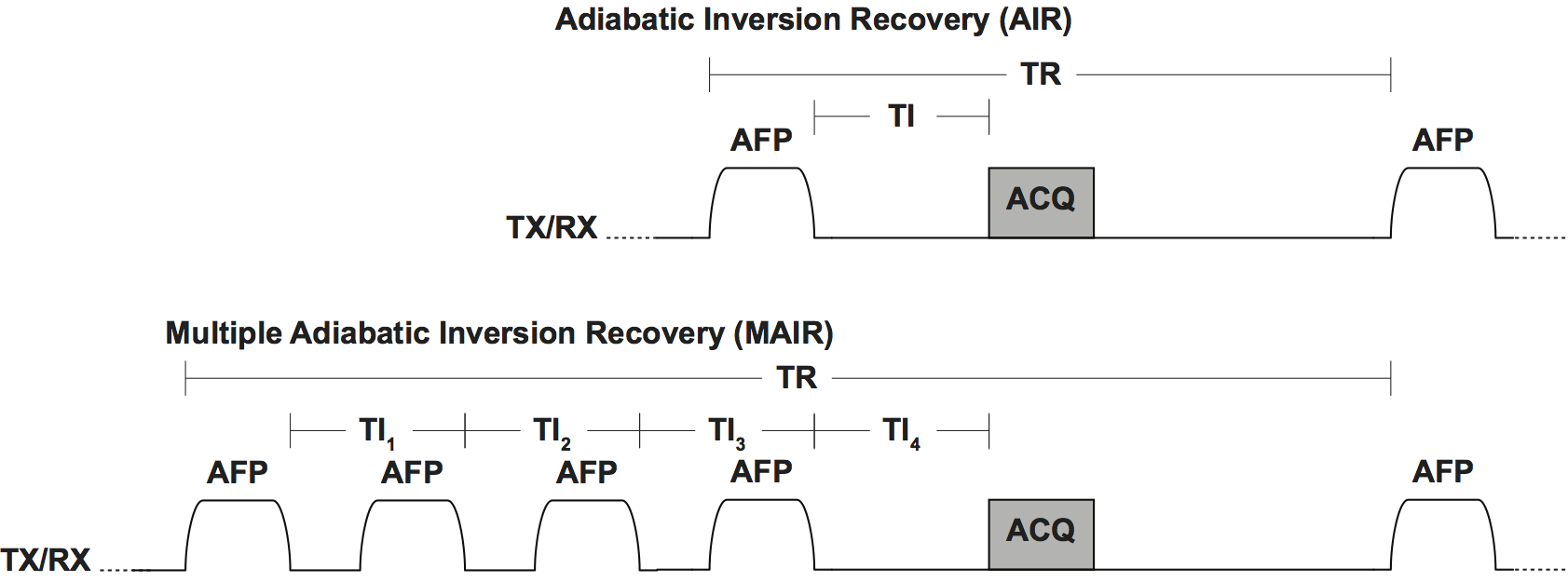
Towards the assessment of individual fracture risk in human bone, the T2 selectivity of adiabatic inversion recovery (AIR) RF pulses are used with 3D ultrashort echo time (UTE) imaging to distinguish individual water components in bone (1-4). AIR pulses suppress short-lived T2 signal from water bound to the collagen matrix in bone (i.e. bound water) and invert free water signal in the bone pore space (i.e. pore water). Bound water can then be imaged independently of pore water with an AIR sequence timed to null the pore water signal. However, reliable suppression of pore water depends on accurate characterization of pore water T1. While population-averaged T1 values haven been used in previous work (5), pathophysiological variations in pore water T1 (6,7) across subjects will result in the contamination of bound-water measurements with unsuppressed pore water signal. We propose to reduce this T1-dependence by extending the AIR sequence to a multiple adiabatic inversion recovery (MAIR, Fig 1) sequence optimized to suppress pore water signal over a broad T1 domain. Note that in clinical practice, the extension of AIR to MAIR will require ~4X increase in TR (and therefore scan time) due to SAR constraints, but MAIR is only practical with 2D UTE, as recently demonstrated for AIR (8).Methods
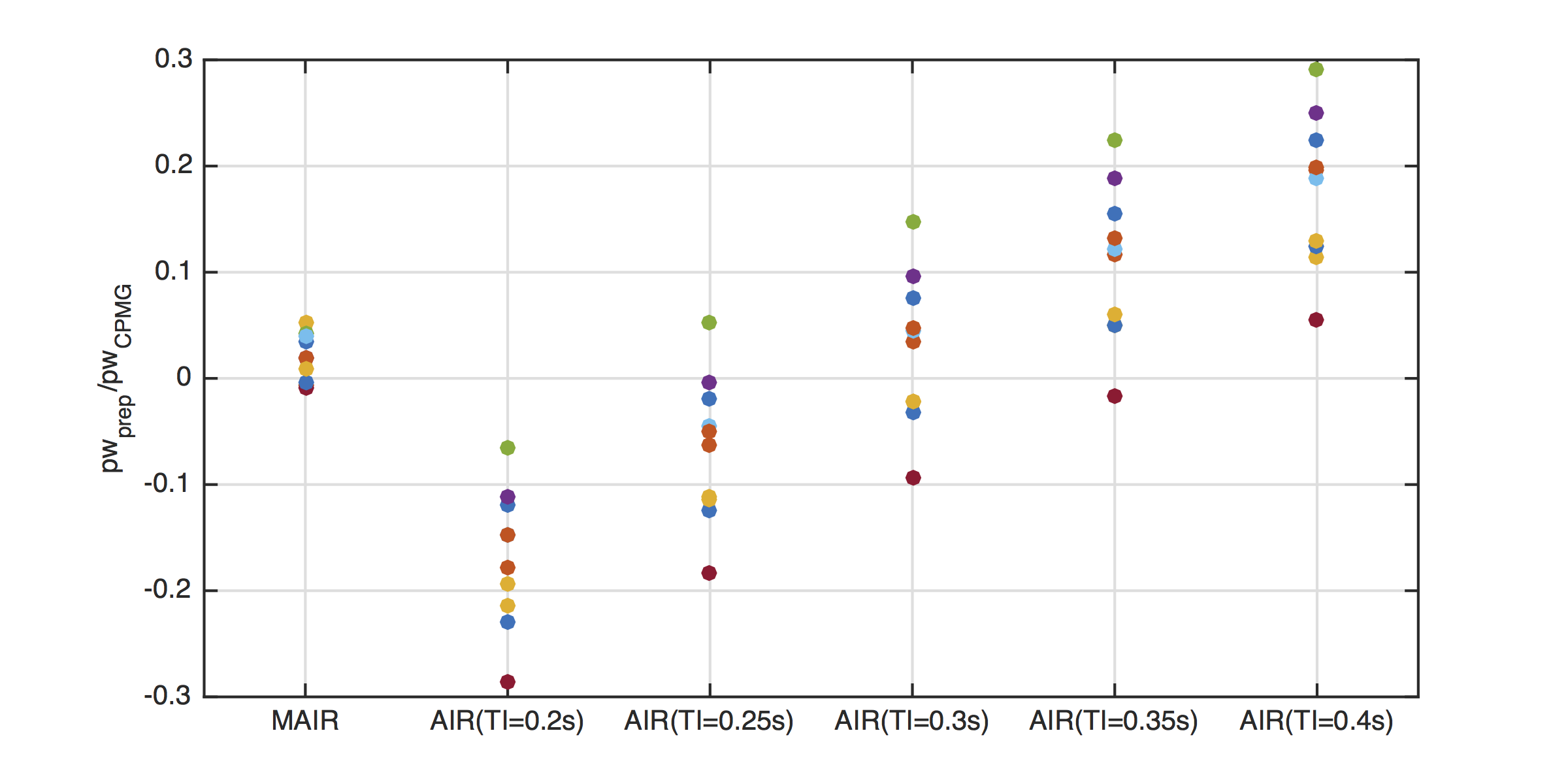
Cortical bone samples were bored from ten cadaver donors. For each sample, Carr-Purcell-Meiboom-Gill (CPMG) T2 measurements were performed on a 4.7T Agilent system with TR = 15 s, TE = 100 µs, and 10,000 total echoes (9). AIR-CPMG measurements were performed in a range of inversion recovery times near the average T1 null point for pore water (TI = 0.2, 0.25, 0.3, 0.35 and 0.4 s). MAIR-CPMG measurements were performed with four adiabatic pulses, as shown in Figure 1. Inversion recovery times were selected to suppress signals across the T1 range 0.2 - 2 s (TI1/2/3/4 = 1.998/0.890/0.398/0.116 s). T2 spectra were calculated from multi-echo data, and pore water signal (pw) was calculated as the sum of all signals with T2 > 1 ms. AIR and MAIR pore water suppression was calculated as pwAIR/pwCPMG and pwMAIR/pwCPMG, respectively.Results and Discussion
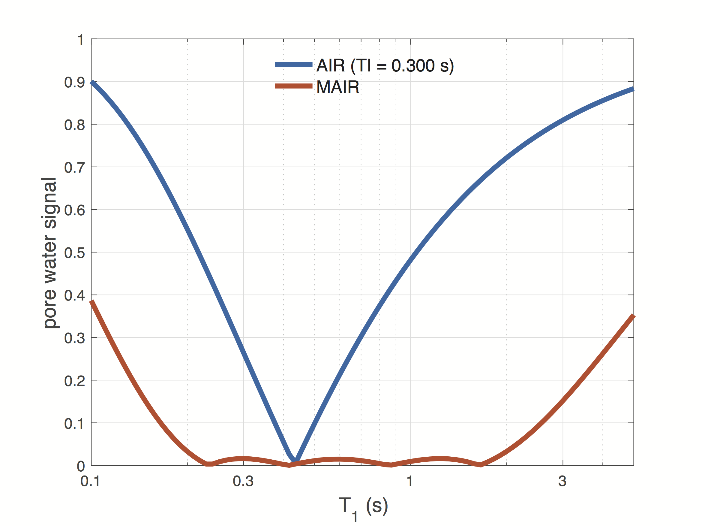
AIR and MAIR signal equation calculations are shown in Figure 2. An AIR TI = 0.3 s results in a nulling of signal exhibiting a T1 ≈ 0.430 s. However, the MAIR sequence provides robust saturation of T1 values between 0.2 and 2 s.
AIR- and MAIR-suppressed pore water signal is shown in Figure 3 for all 10 bone samples. With AIR preparation, an inversion time of about 0.3 ms produces the smallest average contribution of pore water, with a standard deviation of ≈ 2.3%. However, since pore water T1 values vary naturally within the set of samples, the AIR inversion time required to completely null pore water signal also varies. MAIR is able to improve pore water suppression by saturating signal from a broad domain of T1 values, resulting in a standard deviation of < 1%.
Conclusion
Multiple adiabatic inversion preparation can be used to null signals exhibiting a large range in T1 values. For bound water quantification and the assessment of bone fracture risk, this method results in robust suppression of pore water signal without requiring accurate pore water T1 measurement.Acknowledgements
No acknowledgement found.References
1. Larson PEZ, Conolly SM, Pauly JM, Nishimura DG. Using adiabatic inversion pulses for long-T-2 suppression in ultrashort echo time (UTE) imaging. 2007;58(5):952–61.
2. Du J, Bydder M, Takahashi AM, Carl M, Chung CB, Bydder GM. Short T2 contrast with three-dimensional ultrashort echo time imaging. Magn Reson Imaging. 2011 May;29(4):470–82.
3. Horch, R. A., Gochberg, D. F., Nyman, J. S. and Does, M. D. (2012), Clinically compatible MRI strategies for discriminating bound and pore water in cortical bone. Magn Reson Med, 68: 1774–1784. doi:10.1002/mrm.24186
4. Manhard, M. K., Horch, R. A., Gochberg, D. F., Nyman, J. S., Does, M. D., Bone. (2015). In Vivo Quantitative MR Imaging of Bound and Pore Water in. Radiology, 277(1), 221–230. http://doi.org/10.1148/radiol.2015140336
5. Manhard MK, Uppuganti S, Granke M, Gochberg DF, Nyman JS, Does MD. MRI-derived bound and pore water concentrations as predictors of fracture resistance. Bone. 2016 Jun;87:1–10. PMCID: PMC4862893
6. Fantazzini P, Garavaglia C, Guglielmi G. Continuous distribution analysis of marrow 1H magnetic resonance relaxation in bone. Magn Reson Imaging. 2001 Feb;19(2):227–31.
7. Fantazzini P, Garavaglia C, Palombarini M, Brown RJS, Giavaresi G, Giardino R. Analysis of 1H-NMR relaxation time distributions in L1 to L6 rat lumbar vertebrae. Magn Reson Imaging. 2004 Jun;22(5):689–95.
8. Manhard MK, Harkins KD, Gochberg DF, Nyman JS, and Does MD. 30-Second Bound- and Pore-Water Maps of Cortical Bone. Proc ISMRM 2016:676
9. Horch, R. A., Nyman, J. S., Gochberg, D. F., Dortch, R. D., & Does, M. D. (2010). Characterization of 1H NMR signal in human cortical bone for magnetic resonance imaging. Magnetic Resonance in Medicine, 64(3), 680–7. http://doi.org/10.1002/mrm.22459
Figures