1581
DTI imaging application of vastus medialis oblique muscle in recurrent patellar dislocation1Radiology Department of Peking University Third Hospital, Beijing, People's Republic of China, 2Radiology Department of Tsinghua Changgung Hospital, Beijing, People's Republic of China, 3GE Healthcare, MR Research China, Beijing, People's Republic of China
Synopsis
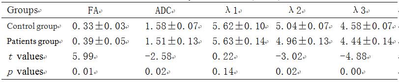
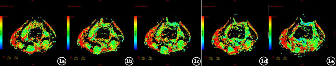
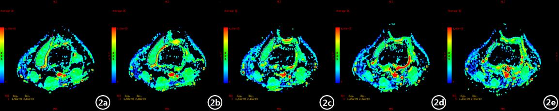
The osseous-related factors that influence patellofemoral joint instability have been well-studied in plenty of the previous reported literatures. However, the muscle-related factors that affect patellofemoral joint instability have not been fully revealed. MRI is a noninvasive imaging method, including conventional MRI scans and diffusion tensor imaging (DTI) sequence, which can respectively reflect the macroscopic and microscopic structures of the muscle fibers.1,2 In the current study, the cross-sectional area were measure with DTI parameters of FA, ADC, and λ1, λ2, λ3 of vastus medialis oblique (VMO). Thereafter, these parameters were compared between the recurrent patellar dislocation patients and the healthy volunteers.
Purpose
The aim of this study was to evaluate the imaging characteristics of vastus medialis oblique (VMO) muscle fibers in the recurrent patellar dislocation using magnetic resonance imaging (MRI) method and to compare the morphology and muscle function of VMO between the recurrent patellar dislocation patients and healthy volunteers.Methods
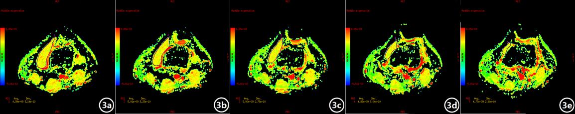
Thirty patients diagnosed with recurrent patellar dislocation and 30 healthy volunteers matched in age, sex and BMI were recruited in the current study. All the subjects underwent conventional MRI scan and DTI imaging. The recurrent dislocation group consisted of 7 males and 23 females (average age 21.37±3.76 years old, average BMI 21.18±3.18kg/m2). On the other hand, the control group consisted of 7 males and 23 females (average age 21.93±3.12 years old, average BMI 20.80±2.64kg/m2). VMO cross-sectional area was derived for both groups using conventional MRI and FA, ADC, λ1, λ2, λ3 values on DTI. t test was used to compare the differences for these six parameters between the recurrent dislocation and control groups.Results
VMO muscle FA, ADC, λ2 and λ3 have better statistical significance in recurrent dislocation group than those of the control group(p < 0.05),while VMO muscle cross-sectional area and λ1 demonstrated no statistical significance between two groups(p > 0.05).Discussion
The recurrent dislocation group demonstrated increased VMO muscle FA, while ADC, λ2 and λ3 reduced significantly (significance<0.05), compared to the control group. These differences in parameters indirectly reflected the shortening of the muscle fiber diameter, which weakened the muscle strength. In addition, the cross-sectional area of VMO exhibited a decreasing trend, but with no statistical difference. The reason may be due to the delay in muscle morphological change while compared to muscle microstructural change. DTI technology could potentially reflect the microstructural changes of the muscles with specific measurements at the early stage of the disease.Conclusion
VMO muscle function is closely associated with recurrent patellar dislocation, where MRI technology, especially DTI, can reflect the modifications of muscle function at the early stage. DTI imaging may be a potential non-invasive assessment tool on monitoring the muscle function status in recurrent patellar dislocation.Acknowledgements
No acknowledgement found.References
1. Balcarek P, Oberthür S, Frosch S, et al. Vastus medialis obliquus muscle morphology in primary and recurrent lateral patellar instability. Biomed Res Int. 2014;2014:326586.
2. Kan JH, Heemskerk AM, Ding Z, et al. DTI-based muscle fiber tracking of the quadriceps mechanism in lateral patellar dislocation. J Magn Reson Imaging. 2009;29(3):663-670.
Figures