1579
A Potential Pathophysiological Link Between Generalized and Localized Muscle Fat Infiltration in Chronic Whiplash Patients1Department of Biomedical Engineering, Linköping University, Linköping, Sweden, 2Center for Medical Image Science and Visualization, Linköping University, Linköping, Sweden, 3Department of Medical and Health Sciences, Physiotherapy, Linköping University, Linköping, Sweden, 4Department of Physical Therapy and Human Movement Sciences, Feinberg School of Medicine, Northwestern University, Chicago, IL, United States, 5Department of Medical and Health Sciences, Linköping University, Linköping, Sweden
Synopsis
Muscle fat infiltration (MFI) in the cervical multifidi muscles and whole-body skeletal muscle tissue was measured in participants with chronic whiplash associated disorders (WAD) using whole-body fat-water separated MRI to investigate potential interaction between deep neck muscle and generalized MFI. Thirty participants with chronic WAD and 30 matched controls were included. MFI in multifidi was strongly associated to whole-body MFI as well as to severity of WAD. The strong association between Multifidi MFI and whole-body MFI indicates that both generalized factors and localized effects related to the trauma may be important for understanding the pathophysiology of chronic WAD.
Purpose
To investigate if muscle fat infiltration (MFI) in the cervical multifidi muscles is associated with MFI in segmented whole body musculoskeletal tissue in participants with chronic Whiplash Associated Disorder (WAD) and matched controls.Methods
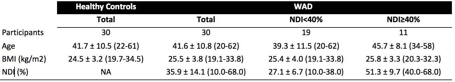
Thirty subjects with mild (N=19) or severe (N=11) chronic WAD (at least 6 months duration) and 30 matched controls were included. The neck disability index (NDI)1 was used for stratification (mild: 20 < NDI < 40 % and severe: NDI ≥ 40 %). See Table 1 for descriptive statistics.
The neck region was imaged using high-resolution fat- and water-separated two-point Dixon imaging in 0.75*0.75*0.75 mm3 isotropic resolution. Whole body fat- and water-separated two-point Dixon images were acquired in lower resolution of 1.75*1.75*1.75 mm3. Both the neck region and the whole body were imaged on a Philips Ingenia 3T scanner using a head coil and anterior and posterior body array coils.
Following water-fat separation by phase sensitive reconstruction2 the right and left m. multifidi were segmented using a semiautomatic segmentation method3 from cervical level C3 to cervical level C7 in the high-resolution dataset and the fat signal fraction (fat/(fat+water)) was calculated. Figure 1 illustrates the segmentation in one participant in three different planes.
Automated segmentation of whole-body muscle tissue using the method described in Karlsson et al 20154 was applied following water-fat separation2 and intensity inhomogeneity correction using fat signal referencing5, 6. MFI was measured in each segmented muscle group by calculation of the average intensity corrected fat signal within each muscle mask.
Mixed linear model analysis was used to investigate the association between MFI in m. multifidi and average whole body MFI measured within segmented muscles from lower and upper legs as well as the abdominal region. In Figure 2 the included muscle regions are shown. Muscles in the arms and the rotator cuff were excluded due to limitations in field of view.
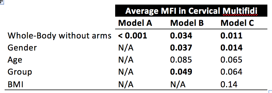
In the model using MFI in m. multifidi as response variable the following effects were included: Model A: whole-body MFI; Model B: whole-body MFI, age, gender, Group (control, WAD (mild), WAD (severe)); Model C: whole-body MFI, age, gender, Group (control, WAD (mild), WAD (severe)), BMI p-values < 0.05 was considered significant.
Results
Model A showed a strongly significant (p<0.001) association between whole body MFI and MFI in m. multifidi (Table 2). The association remained significant in model B when introducing correction for age, gender and WAD group as well as when BMI was included as effect in model C. In model B but not in model C there was a significant association to WAD group. Post hoc tests showed that the participants with severe WAD had significantly higher MFI than participants with mild WAD.Discussion
MFI in m. multifidi is significantly associated with fat infiltration in whole body musculoskeletal tissue. This association holds for correction of the most important confounders in chronic WAD; age, gender and BMI. There was an association between severity of WAD and MFI in m. multifidi also after correction for whole body MFI indicating that the specific MFI of m. multifidi is of particular interest in understanding the pathophysiology of chronic WAD. The association between whole body MFI and MFI in m. multifidi indicates a potential generalized pathophysiological link resulting in elevated whole body MFI in WAD subjects with poor recovery as well as a link directly related to the mechanistic effects of the trauma, which may affect the deep neck muscles as evidenced by elevated levels of multifidi MFI. Further research is needed to clarify whether this generalized component of MFI is present prior to the whiplash trauma or not.Conclusion
The strong association between multifidi MFI and whole-body MFI indicates that both generalized factors and localized effects related to the trauma may be important for understanding the pathophysiology of chronic WAD.Acknowledgements
Purpose The study was funded through the Swedish Medical Research Council and the Medical Research Council of Southeast Sweden.References
1. Vernon H, Mior S. The Neck Disability Index: a study of reliability and validity. J Manipulative Physol Ther. 1991 Sep; 14(7):409-15
2. Romu, T., Dahlström, N., Leinhard, O. D. and Borga, M. (2016), Robust water fat separated dual-echo MRI by phase-sensitive reconstruction. Magn. Reson. Med.. doi:10.1002/mrm.26488
3. Malmberg F, Lindblad J, Nyström I. Sub-pixel segmentation with the image forecasting transform. Playa dle Carmen, Mexico; 2009.
4. Karlsson, A., Rosander, J., Romu, T., Tallberg, J., Grönqvist, A., Borga, M. and Dahlqvist Leinhard, O. (2015), Automatic and quantitative assessment of regional muscle volume by multi-atlas segmentation using whole-body water–fat MRI. J. Magn. Reson. Imaging, 41: 1558–1569. doi:10.1002/jmri.24726
5. Dahlqvist Leinhard O, Johansson A, Rydell J, et al. Quantitative abdominal fat estimation using MRI. In: Proc. Int. Conf. Pattern Recog (ICPR). 2008; art.no 4761764.
6. Romu T, Borga M, Dahlqvist Leinhard O. MANA – multi scale adaptive normalized averaging. In: Proc. Int. Symp. Biomed. Imaging (ISBI) 2011; art.no. 5872424:361-364.
Figures


Figure 2 - A coronal view of the included automatically segmented muscles groups overlaid on a combined fat and water image. The resolution is 1.75*1.75*1.75 mm3.