1575
Fat fraction and muscular volume of entire supraspinatus muscle with/without tendon tear using IDEAL-IQ sequence1Radiology, St.Luke's International Hospital, Tokyo, Japan, 2Department of Intelligent Image Information, Gifu University, 3St.Luke's International Hospital, 4Radiology, University of California, Irvine
Synopsis
Muscular
atrophy and fatty degeneration of the supraspinatus
muscle occurs after rotator cuff tear. It
is often necessary to evaluate for these muscle abnormalities following tears
to determine surgical candidacy. We usually
evaluate this on a single slice. However, it is not well known whether this
slice is representative of fatty degeneration and
muscular atrophy overall.
Furthermore, it is also not
known how fatty degeneration and muscular atrophy will progress
after tear. The purpose of this study was to make a standardized fat fraction and muscular volume model in patients with or without supraspinatus tendon tear using IDEAL-IQ sequence.
Introduction
Muscular atrophy and fatty degeneration of the supraspinatus muscle occurs after rotator cuff tear. It is often necessary to evaluate for these muscle abnormalities following tears to determine surgical candidacy (1). The usefulness of fat fraction measurement of the rotator cuff muscles on preoperative MRI was reported for predicting re-tear of the tendon (2). We usually evaluate this on a single slice, the most lateral oblique slice on a sagittal Y-view (CYV). However, it is not well known whether this slice is representative of fatty degeneration and muscular atrophy overall. Furthermore, it is also not known how fatty degeneration and muscular atrophy will progress after rotator cuff tear. The purpose of this study was to make a standardized supraspinatus muscle fat fraction and muscular volume model in patients with or without tendon tear by using IDEAL-IQ technique on 3T MRI.Materials and methods
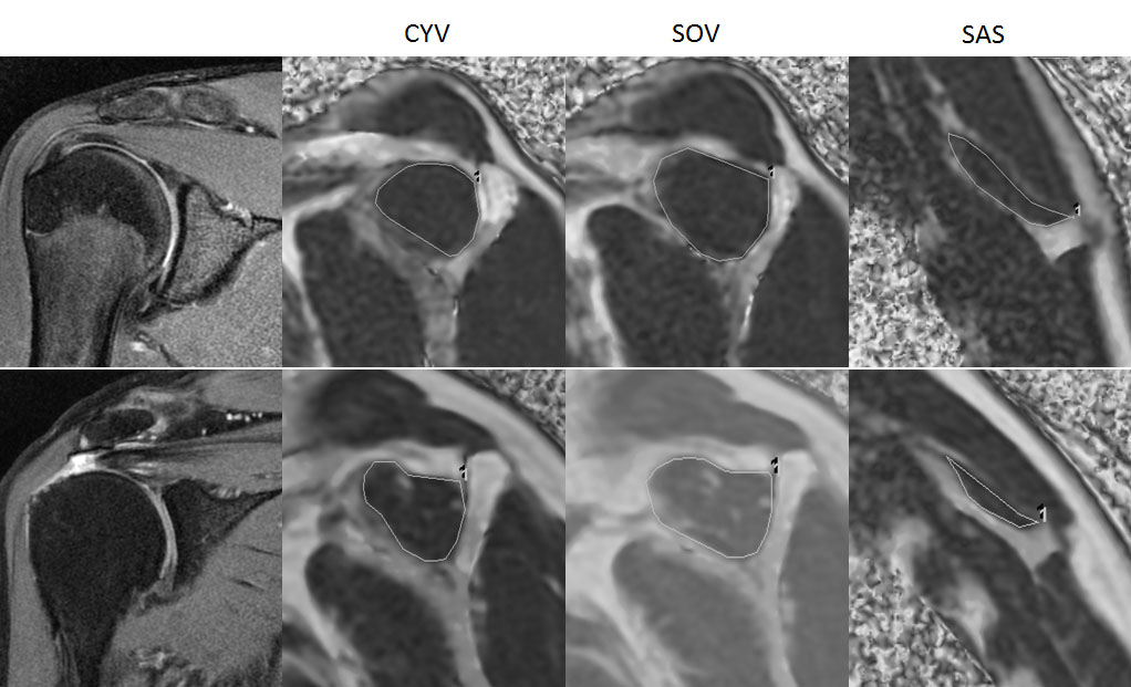
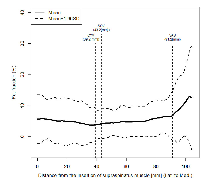
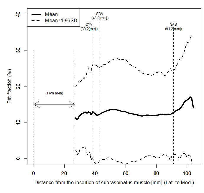
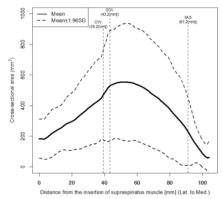
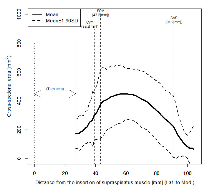
This study included 29 patients with full-thickness rotator cuff tears (17 men, 12 women; mean age, 71.9 ± 6.5 years; age rage, 60-90 years) and 31 patients without tears (17 men, 14 women; mean age, 59.1 ± 15.5 years; age range, 16-87 years). All MR examinations were performed on a 3.0-T unit (Discovery MR750w, GE Healthcare, Barrington, USA) using a 3-channel shoulder array coil. Whole supraspinatus muscle images were obtained using an oblique sagittal IDEAL-IQ sequence, and then fat fraction values and muscular volume were measured on fat fraction images with slice-by-slice manual segmentation by a board-certified radiologist on the PACS system with a region of interest (ROI) placed over supraspinatus muscle (Fig.1). The entire normalized supraspinatus model was created by matching 3 anatomical landmarks (CYV: the conventional Y-view, SOV: the supraspinatus origin-view, SAS: the superior angle of scapula-view) with Lagrange’s interpolation. We calculated the average fat fraction values and muscular volume over the entire normalized muscle with 2D graphs. The differences in average fat fraction values and muscular volume between the full-thickness tear group and non-tear group were analyzed using the Wilcoxon signed-rank test.Results and discussion
In the full-thickness tear group, the average tear length of the supraspinatus tendon was 2.7cm. The fat fraction values over the entire muscle were statistically higher in the full-thickness tear group than non-tear group (12.7% vs 5.7%, p<0.001). Fat fraction values of the supraspinatus muscle were relatively consistent regardless of within muscle measurement location (i.e. fat fraction values were independent of measurement location) (Figs.2 and 3). The muscular volume over the entire muscle was statistically lower in the full-thickness tear group than non-tear group (25.44 ml vs 36.99 ml, p<0.001). The cross-sectional area was greatest in the muscle belly region in both groups, although the muscle belly location in the full-thickness tear group was displaced medially due to retraction (Figs.4 and 5).Conclusions
We applied an IDEAL-IQ sequence for the evaluation of fatty degeneration and muscular volume over the entire supraspinatus muscle in patients with or without supraspinatus tendon tears. The fat fraction within muscle was relatively consistent even in patients with full-thickness rotator cuff tear, while the muscular volume on the cross-sectional area was variable in patients with/without full-thickness rotator cuff tear. Hence the fat fraction value would be feasible and easy to use as an imaging biomarker for clinical applications in preoperative evaluation.Acknowledgements
No acknowledgement found.References
(1) Mellado JM et al. Am J Roentgenol. 2005; 184(5): 1456-63.
(2) Nozaki T et al. Radiology. 2016; 280(2): 500-9.
Figures