1542
Multiparametric MRI assessment reveals early cartilage degeneration at 2 and 8 weeks after anterior cruciate ligament transection in a rabbit model1Research Unit of Medical Imaging, Physics and Technology, University of Oulu, Oulu, Finland, 2Medical Research Center, University of Oulu and Oulu University Hospital, Oulu, Finland, 3Department of Applied Physics, University of Eastern Finland, Kuopio, Finland, 4Diagnostic Imaging Center, Kuopio University Hospital, Kuopio, Finland, 5Human Performance Laboratory, Faculty of Kinesiology, University of Calgary, Calgary, AB, Canada, 6Department of Diagnostic Radiology, Oulu University Hospital, Oulu, Finland
Synopsis
The sensitivity of several quantitative MRI parameters to early degenerative changes in articular cartilage was investigated in an experimentally induced anterior cruciate ligament transection (ACLT) rabbit model. Namely T1, continuous wave T1ρ (CWT1ρ), adiabatic T1ρ (AdT1ρ) and T2ρ (AdT2ρ), T2 and TRAFF (relaxation along a fictitious field) were measured at 9.4 T. All parameters showed significant elongation within two weeks of ACLT, except TRAFF. Rotating frame parameters showed some evidence of superior sensitivity to cartilage alterations. A multiparametric approach may enhance the discrimination power of quantitative MRI for early cartilage degeneration.
PURPOSE
To study the sensitivity to degenerative cartilage changes at two and eight weeks after ACLT1 for several quantitative MRI parameters, including T1, T2, continuous wave T1ρ (CWT1ρ), adiabatic T1ρ (AdT1ρ) and T2ρ (AdT2ρ)2 and TRAFF (relaxation along a fictitious field)3.METHODS
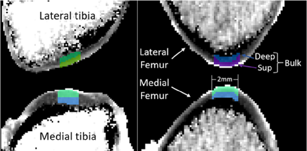
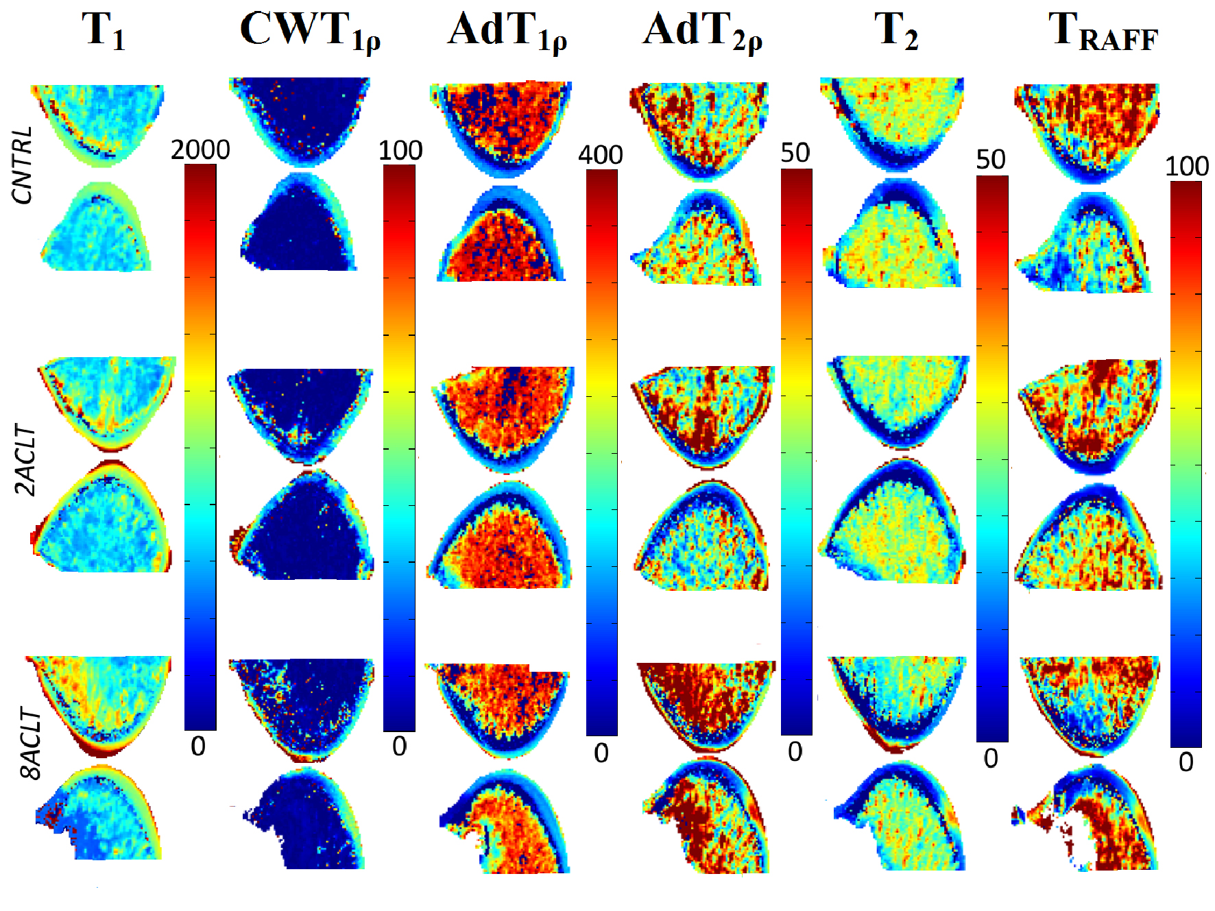
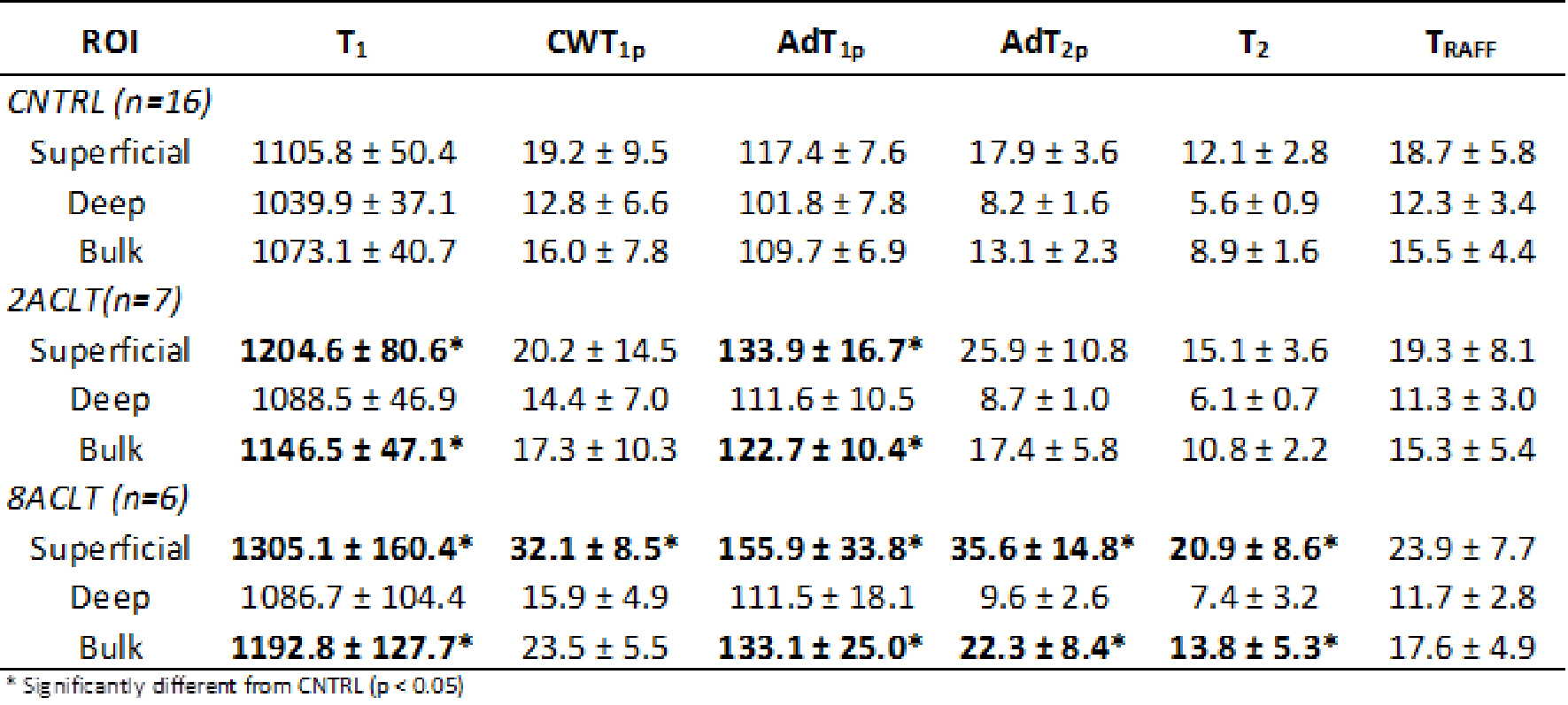
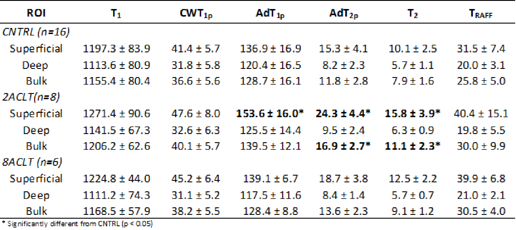
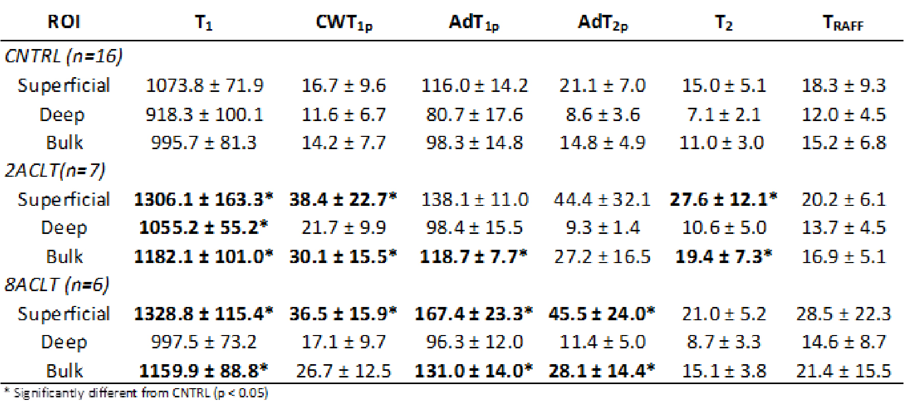
ACLT was surgically induced in the knees of skeletally mature New Zealand White rabbits. The rabbits were 12 months of age at the beginning of the experiment, when unilateral anterior cruciate ligament transection (ACLT) was performed on 14 rabbits. Snapshots of disease progression were captured by sacrificing animals at two weeks (2ACLT, n=8) and eight weeks (8ACLT, n=6) after surgery. An additional 16 non-operated rabbits served as control. Femoral condyles and tibial plateaus were harvested for biomechanical testing and MRI measurements (Fig.1). ACLT surgical operations were conducted according to the guidelines of the Canadian Council on Animal care and were approved by the Animal Ethic committee at the University of Calgary. Specimens were assessed on a 9.4 T MRI scanner (Oxford instruments Plc, Witney, UK) in combination with a 19-mm quadrature RF volume transceiver (RAPID Biomedical GmbH, Rimpar, Germany). The following parameters were measured using different magnetization preparations followed by a fast spin echo readout (TR/TEeff=5s/4.3ms, matrix=256x128, slice thickness=1mm and FOV=18x18mm2): (1) T1 (TI: 200,500,800,1100,1400 and 3000 ms); (2) CWT1ρ (CW spin-lock pulses (γB1=1kHz) of 0,4,8,16,32,64 and 128 ms embedded between AHP pulses); (3) AdT1ρ (trains of 0,4,8,12,24 and 36 adiabatic full-passage (AFP) pulses of 4.5 ms, γB1,max=2.5kHz); (4) AdT2ρ (trains of 0,4,8,12 and 24 AFP pulses between adiabatic half-passage (AHP) pulses of 4.5 ms per pulse, γB1,max=2.5kHz); (5) T2 (TE = 8.7,12.6,18.2,26.4,38.2,55.3 and 80 ms); (6) TRAFF (trains of 0,2,4,8 and 16 RAFF pulses of 9 ms length, γB1,max=625Hz). Maps were calculated for all parameters using MATLAB (Fig.2). The mean values for each parameter were determined for 2-mm wide cartilage ROIs drawn in the load bearing areas of the lateral and medial femur (LF and MF, respectively) and the lateral and medial tibia (LT and MT, respectively) with the articular surface oriented perpendicular with respect to B0 to minimize the magic angle effect (Fig.1). Additionally, laminar analysis was performed and the mean values for each parameter were determined for the superficial 50% and the deep 50% of the cartilage samples. Differences between groups were tested using Kruskal-Wallis post-hoc tests.RESULTS
All MRI parameters showed significant elongation in ACLT groups (Tables 1-3), except for TRAFF. Furthermore, for each parameter, differences were significant at two weeks following surgery in at least one compartment of the knee, consistent with the biomechanical findings: both femoral condyles in ACLT group at two weeks had softer cartilage than controls. Longer T1, CWT1ρ, AdT1ρ, AdT2ρ and T2 were found in degenerated cartilage of superficial and bulk ROI in MF and LF as compared to controls (10-130%). Moreover, the 2ACLT group showed significant elongation of AdT1ρ, AdT2ρ and T2 in MT (Table 2). No significant differences were found in LT (data not shown). No significant differences were observed between 2ACLT and 8ACLT groups in any compartment.DISCUSSION
ACLT in rabbits provides a well-established experimental model for early cartilage degeneration. Previously, elongated values of cartilage CWT1ρ, AdT1ρ, AdT2ρ and T2 in an ACLT rabbit model have been reported at four weeks post surgery4. In the present study, the same quantitative MRI parameters could identify cartilage degeneration as early as two weeks after ACLT. In terms of relative differences in mean values, CWT1ρ and AdT2ρ were the most sensitive parameters and changed by 130% and 116% compared to control knees, respectively. AdT1ρ was the only parameter showing significant elevation in three different compartments within two weeks post surgery. Although the relative differences in mean values were smaller compared to other parameters, longer T1 values were observed in femur two weeks after ACLT also in the deep cartilage. TRAFF did not show any significant differences, consistent with a previous study4. Differences were mostly significant in the superficial cartilage, consistent with the hypothesis that degeneration starts from the most superficial layers5. Regional differences in MRI parameters at different time intervals from ACLT suggest that a multi-parametric approach may provide a more comprehensive assessment of cartilage tissue status at different degrees of degeneration than a single parameter approach.CONCLUSIONS
MRI parameters can distinguish
cartilage changes in an ACLT rabbit model of cartilage degeneration as early as two
weeks post surgery and reflect biomechanical findings. Rotating frame
parameters showed some evidence of superior sensitivity to cartilage alterations. A
multi-parametric approach may enhance the discrimination power of quantitative
MRI for osteoarthritis progression.Acknowledgements
Support from the Academy of Finland (grants #285909 and #293970) and Jane and Aatos Erkko Foundation is gratefully acknowledged.References
1. Nelson et al. OsteoArth Cart 14:114-119, 2006.
2. Michaeli et al. J Magn Reson 169:293-299, 2004.
3. Liimatainen et al. Magn Reson Med 64: 983-994, 2010.
4. Rautiainen et al. OsteoArth Cart 22(10):1444-1452, 2014.
5. Mosher et al. Arthritis & Rheum 50(9):2820-2828, 2004.
Figures