1537
Accelerated Musculoskeletal MRI using Region of Interest Compressed SensingAmaresha Shridhar Konar1,2, Nithin N Vajuvalli1, Shivaprasad Chikop1, and Sairam Geethanath1
1Medical Imaging Research Center, Dayananda Sagar Institutions, Bangalore, India, 2Healthcare, Wipro GE Healthcare, Bangalore, India
Synopsis
MusculoSkeletal (MSK) MRI is used for analyzing injuries, and the knee injury is one of the common injuries reported in sports. Current work uses Compressed Sensing (CS) based reconstruction technique called Region Of Interest Compressed Sensing (ROICS). The proposed method has been demonstrated on five datasets at chosen acceleration factors. The reconstructed images are compared with the reference images using line intensity profile and NRMSE graph to demonstrate the utility of the proposed ROICS method on MSK MRI. The qualitative and quantitative result shows that ROICS performs better than CS. Current and future work involves the prospective implementation of ROICS on MSK MRI.
Purpose
MusculoSkeletal (MSK) MRI is used for analyzing injuries and tumor and it provides good quality images when there is no motion. A knee injury is one of the common sports injuries and MRI is one of the imaging modality used to analyze1. During anxious or in severe pain it is difficult to lie still during imaging and hence there is a requirement for reduction in the data acquisition and reconstruction. Current work uses Compressed Sensing (CS) 2 based reconstruction technique called Region Of Interest Compressed Sensing (ROICS) 3, 4 to retrospectively reconstruct the MSK-MRI undersampled k-space data to achieve acceleration.Theory
CS is based on data sparsity and data consistency, hence sparser the data, better would be the CS reconstruction. ROICS aims to achieve superior CS performance by confining CS reconstruction to a Region Of Interest (ROI), thereby further increasing the acceleration. This reconstruction technique makes use of a mask with all 1’s inside the ROI and 0’s outside it, the inclusion of this mask as weighting function increases data sparsity resulting in superior performance than CS. Conventional CS reconstruction algorithm can be represented by equation: minm (|| FU(m) – y ||2) + λ || ψ(m) ||1) (1), where, m is the current estimate of the image to be obtained, Fu is the undersampled orthonormal Fourier operator: F(.)* undersampling mask, y is the undersampled k-space measured by the acquisition process, λ is the regularization factor, determined by methods such as Tikhonov regularization or L-curve optimization, Ψ is the sparsifying transform operator and ||.||k is the k-norm operator. Data consistency term was evaluated in the spatial domain and ROICS equation was derived by weighting the spatial data consistency term over a ROI and this results in equation (2): minm (|| F-1(Fu(m) - y) * W ||2 + λ || ψ (m * W)||1) (2), where, F-1 is the inverse Fourier transform and W is the Ns*Ns diagonal matrix containing a spatial weighting that can be used to specify and evaluate a ROI, of the dimensions of the image. Data sparsity is the key criterion in CS and The inclusion of ROI mask acts as a weighting function which restricts the objectives of CS to the ROI thereby increasing sparsity whereas conventional CS reconstruction method was performed for the entire image.Methods
ROICS technique has been applied retrospectively on MSK-MRI data acquired using a 1.5 T scanner, in sagittal orientation using knee coil with the following acquisition parameters: Sagittal Proton Density (PD) Fast Spin Echo (FSE) based sequence with TR/TE :1000/39 ms, FA:160 degrees, multi-slice images with matrix size:320x320. ROI was drawn on a selected slice as shown in Figure 1(a) outlined in yellow to cover the knee region such as articular cartilage, Meniscus, lateral collateral ligament. Figure 1(d) is selected slice to cover ACL, PCL, and patella. Figure 1(b) and 1(e) are the ROI selected region for two represented frames. Figure 1(c) and 1(f) are the line intensity profiles plotted for the line marked in yellow in Figure 1(b) and (e) respectively. Both conventional CS and ROICS technique have been applied to five datasets at chosen acceleration factors of 2x, 3x, 5x, 8x, and 10x to demonstrate the utility of the proposed ROICS method on knee MSK MRI. To find the reconstruction error, Normalized Root Mean Square Error (NRMSE) values, and the line intensity profiles plotted for both CS and ROICS reconstructed images.Results
Figure 2 and Figure 3 shows the images reconstructed retrospectively using CS and ROICS reconstruction methods at chosen acceleration factors. This indicates that at higher acceleration factor (3x and above) ROICS performs better than conventional CS technique and also CS images shows hyperintensity. Line intensity profile plotted for both the methods at chosen acceleration factors reveal that ROICS maintains the consistency in the line intensity profile as compared to CS method. Quantitative evaluation has been performed by calculating NRMSE for both CS and ROICS only for the ROI selected region. Graph (Figure 4) shows that as the acceleration increases error in CS increases and ROICS is showing consistency even at high acceleration factor of 10x.Discussion and Conclusion
ROI can be restricted to the region enclosing articular cartilage, lateral meniscus, synovial fluid, ACL, PCL which would result in better reconstruction and it can be used for the assessment and analysis of the knee injuries. ROICS reconstruction can be further improved by considering arbitrary k-space trajectories. Current and future work involves the prospective implementation of ROICS on MSK MRI.Acknowledgements
No acknowledgement found.References
[1] Majewski, M., Habelt Susanne, and Steinbrück Klaus. "Epidemiology of athletic knee injuries: A 10-year study." The knee 13, no. 3 (2006): 184-188 [2] Lustig, Michael, David Donoho, and John M. Pauly. "Sparse MRI: The application of compressed sensing for rapid MR imaging." Magnetic resonance in medicine 58, no. 6 (2007): 1182-1195. [3] Amaresha Shridhar Konar et al., p.3801, ISMRM 2013, [4] Konar, Amaresha Shridhar, Jain A. Divyaa, Shamshia Tabassum, Rajagopalan Sundaresan, Julianna Czum, Barjor Gimi, Ramesh DR Babu, and Sairam Geethanatha. "Region of interest compressed sensing MRI." Journal of the Indian Institute of Science 94, no. 4 (2014): 407-414.Figures
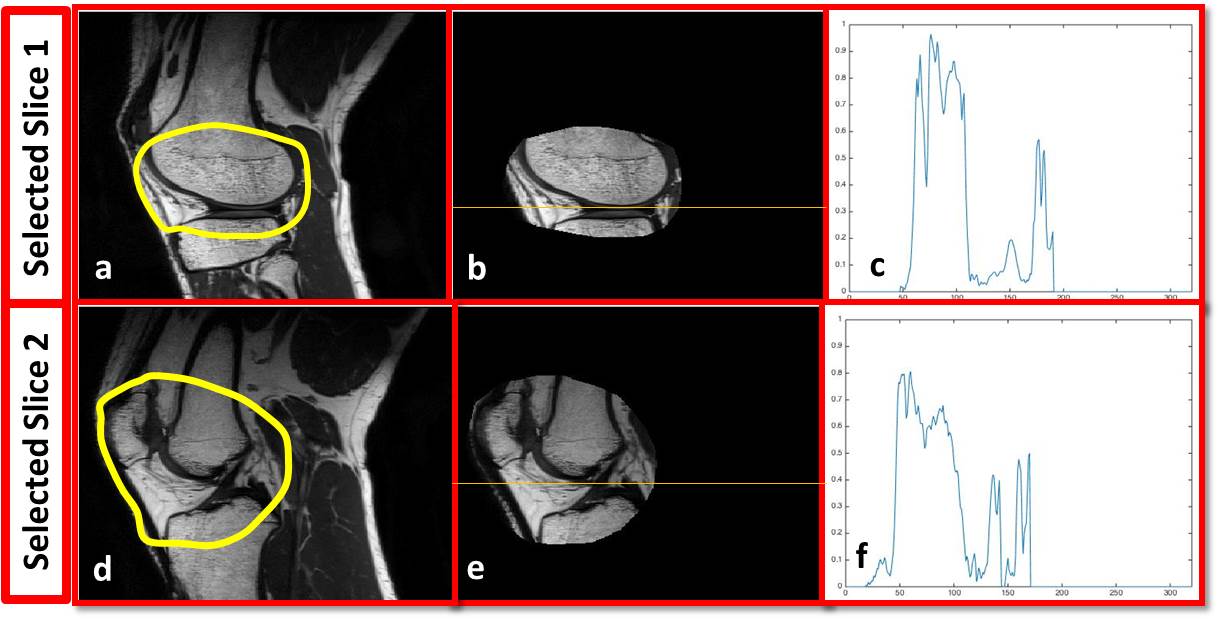
Figure 1: Representative frames: (a) and (d)
shows ROI selected in yellow outline, (b) and (e) ROI masked image, (c) and (f)
are the line intensity profile for the ROI masked reference image shown in
yellow line
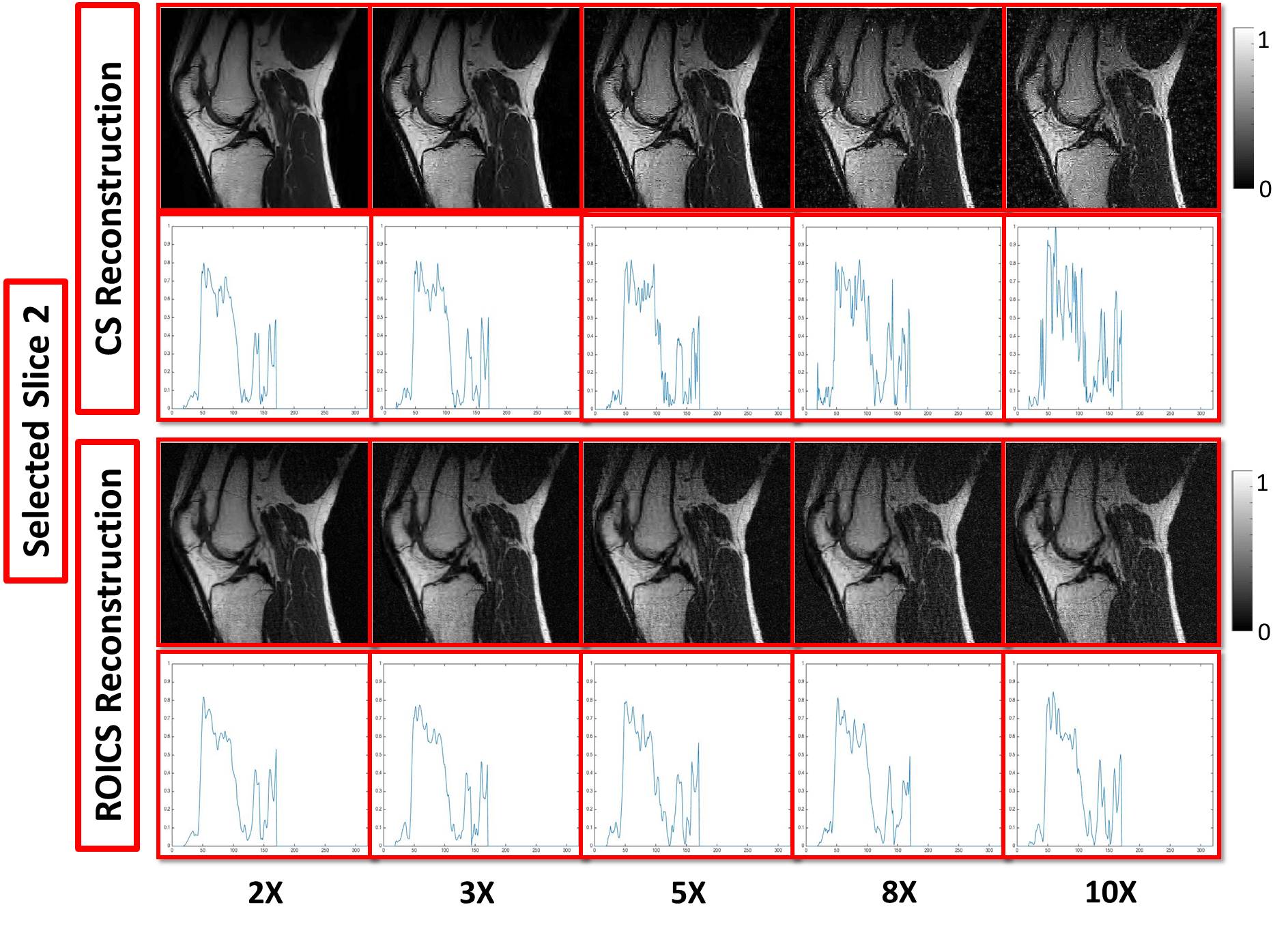
Figure 2: Demonstration of conventional CS and
ROICS reconstruction at chosen acceleration factors with line intensity profile
for the first selected frame.
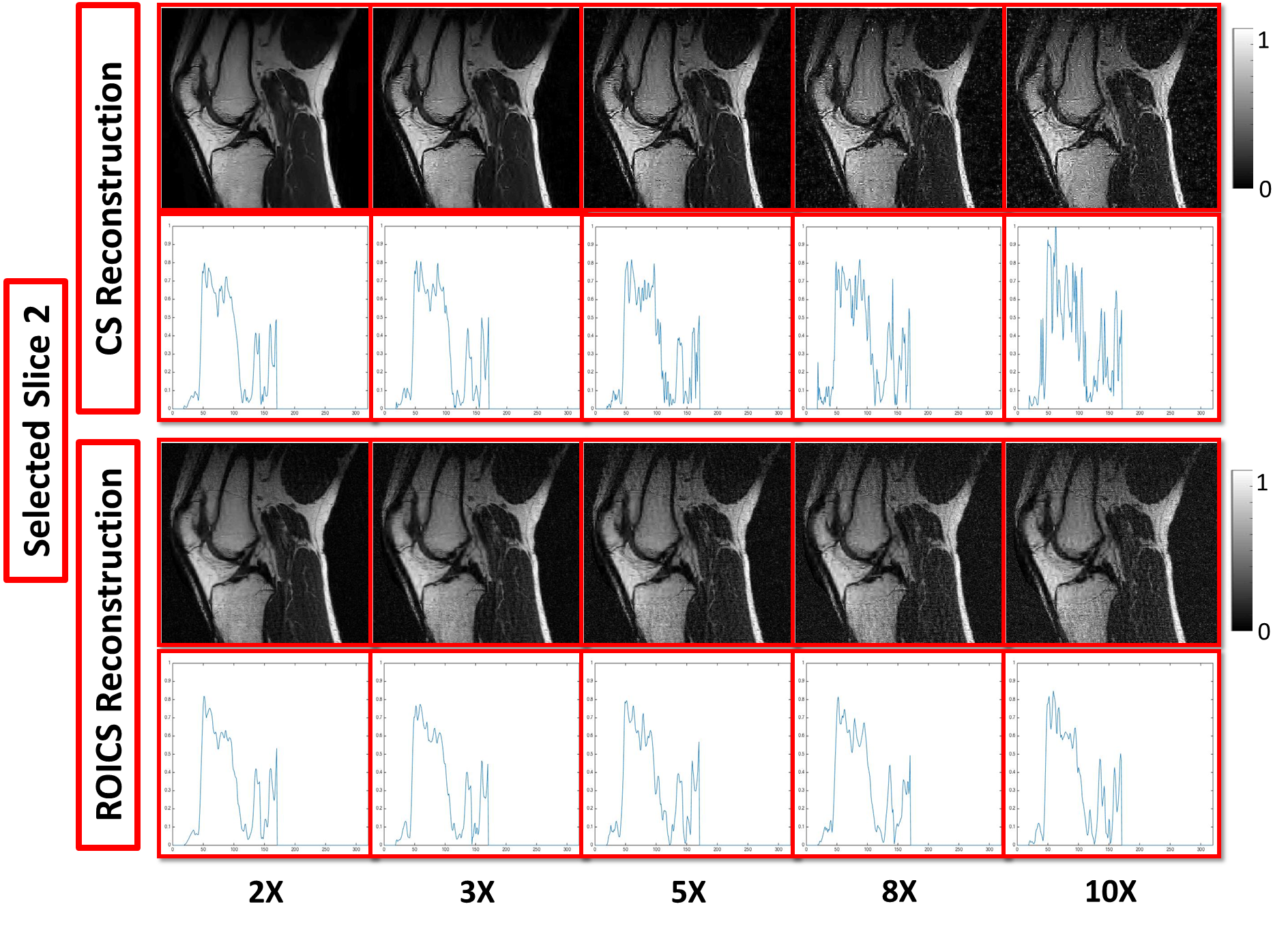
Figure 3: Demonstration of conventional CS and
ROICS reconstruction at chosen acceleration factors with line intensity profile
for the second selected frame.
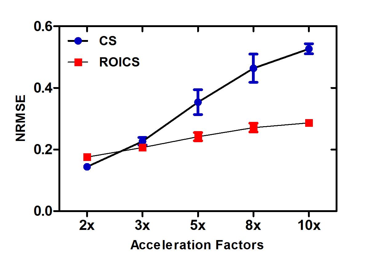
Figure 4: NRMSE comparison between CS and ROICS
method.