1525
Distortion Correction in Readout-Segmented EPI using View Angle Tilting Combined with Phase Modulated RF Pulse1Siemens Shenzhen Magnetic Resonance Ltd, Shenzhen, People's Republic of China
Synopsis
The VAT technique has been applied in ss-EPI to eliminate the distortion along the phase encoding direction. However the long echo spacing in ss-EPI will lead to more phase errors introduced by B0 inhomogeneity and require more VAT gradient, which results an increasing image blurring and limits the spatial resolution. The rs-EPI features much shorter echo spacing compared with ss-EPI, which could be much suitable for VAT. In this study, the VAT technique is integrated into a rs-EPI sequence to further improve the distortion. In addition, phase modulated pulse is used to reduce the image blurring caused by VAT.
Purpose
Readout-segmented Echo Planar Imaging (rs-EPI) with 2D navigation is an established clinical technique for acquiring diffusion-weighted images with a low level of distortion and T2*-related blurring1. Compared with single shot EPI (ss-EPI), the geometric distortions due to susceptibility artifacts could be significantly reduced in rs-EPI. However, for the regions with complex geometry and several susceptibility interfaces (e.g. neck region), the remaining image distortion is still obvious.
The view angle tilting (VAT) technique was initially proposed to correct image distortion caused by B0 inhomogeneity and chemical shift along the readout direction and has been applied in conventional SE sequence2 and then extended to ss-EPI3. However the long echo spacing in ss-EPI will lead to more phase errors introduced by B0 inhomogeneity and require more VAT gradient to correct the distortion, which results an increasing image blurring. It will limit the spatial resolution and consequently make the resolution a trade-off with image blurring. The rs-EPI sequence features much shorter echo spacing compared with ss-EPI, which could be much suitable for VAT, especially for higher resolution acquisitions. In this study, we proposed a new distortion correction method for rs-EPI, in which the VAT technique combined with a phase modulated RF pulse is used to eliminate the distortion caused by the B0 inhomogeneity.
Methods
When VAT is applied to EPI sequence to address the imaging distortion along the phase encoding (PE) direction, the signal S could be expressed as follows: $$s(t_{m}, t_{n})=\int_{x}^{} \int_{y}^{} \int_{\triangle sl + \triangle z(x,y) }^{} \rho(x,y,z)exp(-j\gamma nG_{vat}t_{b}z)\times exp(-j\gamma \triangle B(x,y)nT_{esp})\\\times exp[-j\gamma (mG_{x}\triangle t_{x}x + nG_{y}t_{b}y)]d_{x}d_{y}d_{z}$$ where ΔB(x,y) is the B0 offset on a position coordinate (x, y); GVAT is the VAT gradient applied along the slice selective (SS) direction simultaneously with the PE gradient blips Gy; ρ(x,y,z) is the spin density; γ is the gyromagnetic ratio; Δtx is the dwell time in readout (RO) direction; Tesp is the echo spacing; tb is the blip duration; Δz(x,y) is the position displacement due to B0 inhomogeneity along the SS direction, Δz(x,y) = -ΔB(x,y)/Gz; Gz is the SS gradient; Δsl is the slice thickness; m,n are k-space indices along the RO and PE directions. Assuming GVAT = GzTesp/tb, then the additional phase term caused by B0 inhomogeneity could be completely eliminated. However the addition of the VAT gradient will lead to the blurring along the PE direction. It can be deduced from the above equation that the Fourier transform of the excitation RF slice profile is multiplied along the ky direction as a k-space weighting3.
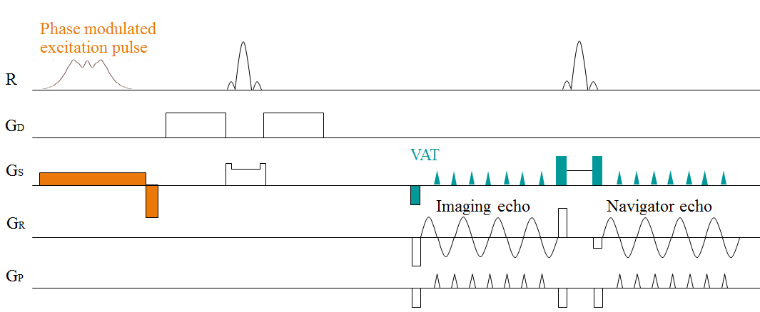
In this study, the VAT technique is integrated into a commercial rs-EPI sequence (RESOLVE, Siemens Healthineers), referred as RESOLVE-VAT. The sequence diagram is illustrated in Fig.1. In addition, a specially designed RF pulse which has a more uniform magnitude modulation function could effectively improve the blurring problem3-4. Here we designed a quadratic RF excitation pulse (duration 22ms, time bandwidth product 20) using the Shinnar-Le Roux (SLR) transformation5-6, in which the modulated RF pulse with minimal B1max is selected as the one with more uniformly k-space weighting along the PE direction. The main lobe width of the pulse is matched to the PE duration, leading to less low-pass filtering effect.
Experiments and Results
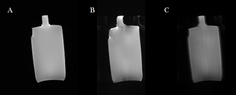
All measurements were performed on a commercial 3T scanner (MAGNETOM Spectra, Siemens Healthineers). Experiment data were obtained from a phantom using a non-product, RESOLVE-VAT sequence. The imaging parameters were as follows: FOV = 350x175mm2, 8 slices with 6mm thickness, matrix = 192x96, echo spacing = 0.34ms, diffusion mode = 4-scan-trace, b = 0, 1000 s/mm2, in-plane GRAPPA factor = 3. For product RESOLVE: TE/TR = 62/3000ms; for RESOLVE-VAT: TE/TR = 89/3000ms. An additional T2-weighted TSE scan was used as a reference. A comparison of sample images from product RESOLVE, RESOLEVE-VAT and TSE is shown in Fig. 2. After the addition of VAT combined with a phase modulated RF pulse, the distortion could be significantly improved and the image structure is more comparable to the one from TSE sequence.Discussion and Conclusion
We have demonstrated that the VAT technique could be integrated into RESOLVE sequence to further improve the distortion problem and the introduction of phase modulated pulse will reduce the image blurring caused by VAT, which is promising clinical applications with high resolution acquisitions. It notes that the phase modulated RF pulse will result in a loss of SNR because of intravoxel phase dispersion cross the slice4. In addition, the long duration of excitation pulse will increase the echo time and further reduce the SNR. More delicate RF design will be investigated to address this problem in the future.Acknowledgements
No acknowledgement found.References
1. Porter D et al. High Resolution Diffusion-Weighted Imaging Using Readout-Segmented Echo-Planar Imaging, Parallel Imaging and a Two-Dimensional Navigator-Based Reacquisition, Magn Reson Imaging. 2009; 62:468-475
2. Cho ZH et al. Total inhomogeneity correction including chemical shifts and susceptibility by view angle tilting. Med Phys. 1988;15(1):7–11
3. Sinyeob A et al. View Angle Tilting Echo Planar Imaging for Distortion Correction. Magn Reson Med. 2012; 68:1211-1219
4. Butts K et al. Reduction of Blurring in View Angle Tilting MRI. Magn Reson Med. 2005; 53:418–424
5. Pauly JM et al. Parameter Relations for the Shinnar-Le Roux Selective Excitation Pulse Design Algorithm. IEEE Trans Med Imaging.1991; 10:53-65
6. Schulte RF et cl. Equi-Ripple Design of Quadratic Phase RF Pulses. J Magn Reson. 2004;166 :111-122
Figures