1524
Pseudo-inverse constrained (PICO) reconstruction reduces colored noise of PROPELLER and improves the gray-white matter differentiation1Graduate Institute of Biomedical Electronics and Bioinformatics, National Taiwan University, Taipei, Taiwan, 2Department of Radiology, University of Cambridge, Cambridge, United Kingdom, 3Department of Applied Physics, National Chengchi University, Taipei, Taiwan, 4Department of Diagnostic Radiology, Li Ka Shing Faculty of Medicine, The University of Hong Kong, Hong Kong, Hong Kong, 5Department of Electrical Engineering, National Taiwan University, Taipei, Taiwan, 6Department of Medical Imaging, Madou Sinlau Hospital, Tainan, Taiwan, 7Department of Medical Imaging, National Taiwan University Hospital, Taipei, Taiwan, 8Department of Medical Imaging, National Taiwan University Hospital Yun-Lin Branch, Taipei, Taiwan, 9Department of Radiology, Cambridge University Hospitals NHS Foundation Trust, Cambridge, United Kingdom, 10MRIS unit, Cambridge University Hospitals NHS Foundation Trust, Cambridge, United Kingdom
Synopsis
The image quality of Periodically Rotated Overlapping ParallEL Lines with Enhanced Reconstruction (PROPELLER) MRI is degraded by the “colored noise” or “blue noise”. Total variation (TV) denoising is expected to reduce colored noise and improve the overall image quality. However, no study has compared different TV algorithms for reducing colored noise. This study explores two TV denoising methods: (1) image domain denoising (IDD), and (2) Pseudo-Inverse
Purpose
Periodically Rotated Overlapping ParallEL Lines with Enhanced Reconstruction (PROPELLER) MR technique provides high-resolution MRI with reduced motion artifacts1. However, the PROPELLER sampling pattern (see Fig. 1(A)) causes the unique “colored noise” or “blue noise”2, which exhibits a higher noise amplitude in the peripheral region of k-space (see Fig. 1(B, C)). While image processing is a routine procedure to reduce image noise, image denoising methods face a trade-off between lowering the noise level or preserving image details.
Edge-preserving total-variation (TV) regularization has recently become a successful method for compressed sensing and image denoising3, but TV denoising has never been validated for reducing colored noise in PROPELLER. The purpose of this study is to test two TV denoising methods for reducing colored noise. Specifically, we considered the following two algorithms in this study: (1) image domain denoising (IDD) method, and (2) our previous Pseudo-Inverse COnstrained (PICO) reconstruction. The image quality of in vivo brain images was scored by radiologists.
Methods
The “edge-preserving” property of TV denoising is expected to preserve the image sharpness and generate piecewise smoothness. Still, the actual image quality is subject to the implementation of each algorithm. The differences between IDD and PICO algorithms are summarized in Table 1. IDD is a fast image domain processing which updates each pixel by the adjacent pixels using Gauss-Seidel model. PICO algorithm is the iterative pseudo-inverse constrained reconstruction, so the k-space trajectory is included in the reconstruction process. IDD in this study uses the scikit-image python module. PICO is based on the python non-uniform fast Fourier transform (pynufft) class. This pynufft module enables multi-coil reconstruction with compute unified device architecture denoising (CUDA) on a graphic processing unit (GPU).
$$$T_2$$$-weighted PROPELLER images were acquired from a 1.5 T scanner (Discovery MR450 1.5T MRI, GE Healthcare, Waukesha, WI, USA) and a 3T scanner (Magnetom Skyra, Siemens AG, Erlangen, Germany). Scanning parameters are TR = 2.5s, TE = 84 - 108 ms, matrix size = 384 × 384 - 512 × 512. Blade width is 24. Sixteen raw data sets were acquired and reconstructed by the conventional PROPELLER reconstruction and two TV denoising algorithms. Three radiologists evaluated the image quality of three different reconstruction methods.
Results
High-resolution PROPELLER images are depicted in Fig. 2. Colored noise is clearly seen in conventional PROPELLER reconstruction (Fig. 2(A)). Both IDD (Fig. 2(B)) and PICO(Fig. 2(C)) reduced this colored noise. However, IDD causes severe image blurring (Fig. 2(B)) and degrades the image details. PICO reduces the image noise and still maintains the image details.
Fig. 3 compares the noise power spectra of three reconstruction methods. IDD fails to reduce the noise in the peripheral region of k-space. PICO generates a more homogeneous k-space noise. Subjective evaluations by three radiologists were listed in Table 2. IDD reduces the image noise but worsens image blurring and gray-white matter differentiation. PICO significantly reduces noise level and increases gray-white matter differentiation. While IDD significantly decreases the overall quality, PICO marginally increases the overall quality.
Discussion
This study implemented two TV denoising algorithms to address PROPELLER related colored noise. Comparing these two algorithms, PICO not only reduces the colored noise in PROPELLER but also preserves image details. In comparison, IDD provides suboptimal image quality and causes significant image blurring. The reasons for the suboptimal quality of IDD are unknown, but it may be related to the unique PROPELLER k-space and colored noise.
Recently, PROPELLER MRI has been used in abdominal MRI4, musculoskeletal MRI5, diffusion imaging6 and chest imaging7. Given the broad applications of PROPELLER MRI, PICO reconstruction can benefit the image quality of clinical PROPELLER MRI.
Conclusion
PICO not only reduces the noise level, but also significantly improves the gray-white matter differentiation and marginally increases the overall image quality of PROPELLER.Acknowledgements
The study was supported by Addenbrooke’s Charitable Trust and the NIHR Comprehensive Biomedical Research Centre award to Cambridge University Hospitals NHS Foundation Trust, in partnership with the University of Cambridge. J.M.L. and H.W.C. received support from the Ministry of Science and Technology under grant MOST 105-2221-E-002-142-MY3.References
1. James G Pipe et al. Motion correction with PROPELLER MRI: application to head motion and free-breathing cardiac imaging. Magnetic Resonance in Medicine, 42(5):963–969, 1999.
2. Fu-Nien Wang, Teng-Yi Huang, Fa-Hsuan Lin, Tzu-Chao Chuang, Nan-Kuei Chen, Hsiao-WenChung, Cheng-Yu Chen, and Kenneth K Kwong. PROPELLER EPI: an MRI technique suitable for diffusion tensor imaging at high field strength with reduced geometric distortions. MagneticResonance in Medicine, 54(5):1232–1240, 2005.
3. Tom Goldstein and Stanley Osher. The split Bregman method for L1-regularized problems.SIAM Journal on Imaging Sciences, 2(2):323–343, 2009.
4. Kyung A Kang, Young Kon Kim, EunJu Kim, Woo Kyoung Jeong, Dongil Choi, Won Jae Lee,Sin-Ho Jung, and Sun-Young Baek. T2-weighted liver MRI using the multivane technique at 3T: Comparison with conventional T2-weighted MRI. Korean Journal of Radiology, 16(5):1038–1046, 2015.
5. Kazuya Nagatomo, Hidetake Yabuuchi, Yuzo Yamasaki, Hiroshi Narita, Seiji Kumazawa,Tsukasa Kojima, Noriyuki Sakai, Masahumi Masaki, and Hiroshi Kimura. Efficacy of periodically rotated overlapping parallel lines with enhanced reconstruction PROPELLER for shoulder magnetic resonance MR imaging. European Journal of Radiology, 85(10):1735–1743,2016.
6. Jameel Muzaffar, Christopher Metcalfe, Steve Colley, and Christopher Coulson. Diffusion-weighted magnetic resonance imaging for residual and recurrent cholesteatoma: A systematic review & meta-analysis. Clinical Otolaryngology, 2016.
7. Pierluigi Ciet, Goffredo Serra, Silvia Bertolo, Sandra Spronk, Mirco Ros, Francesco Fraioli,Serena Quattrucci, M Baroukh Assael, Carlo Catalano, Fabio Pomerri, et al. Assessment of CF lung disease using motion corrected PROPELLER MRI: a comparison with CT. EuropeanRadiology, 26(3):780–787, 2016.
Figures
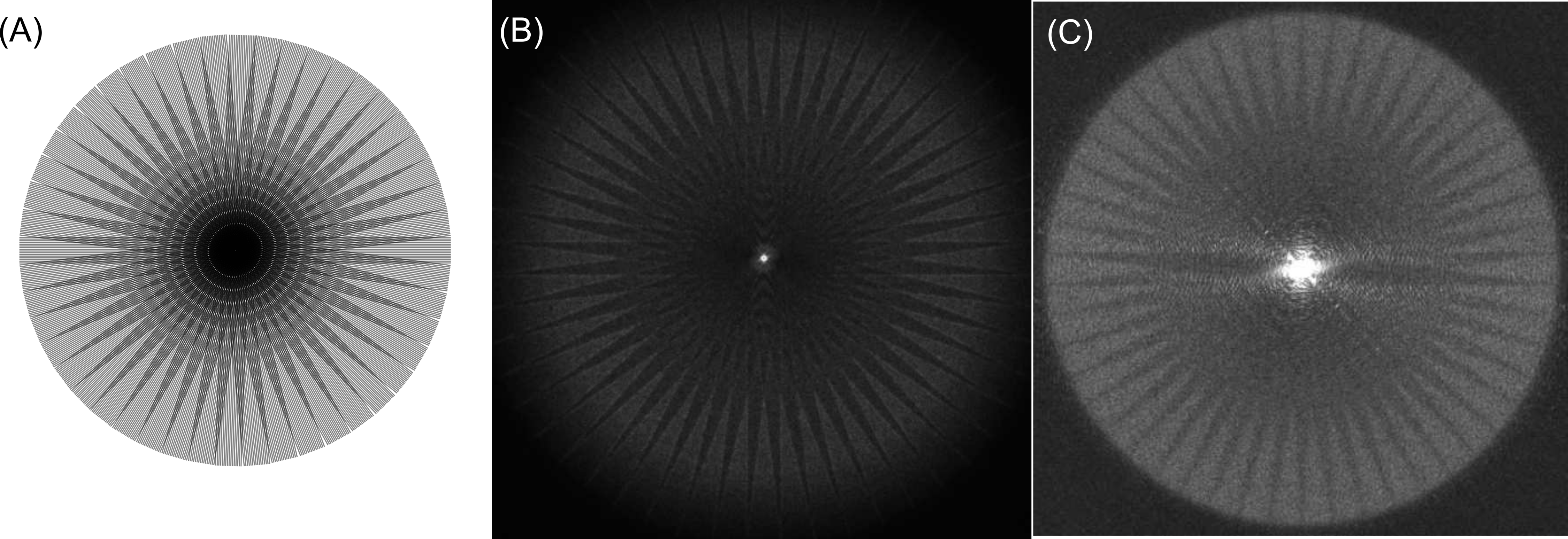
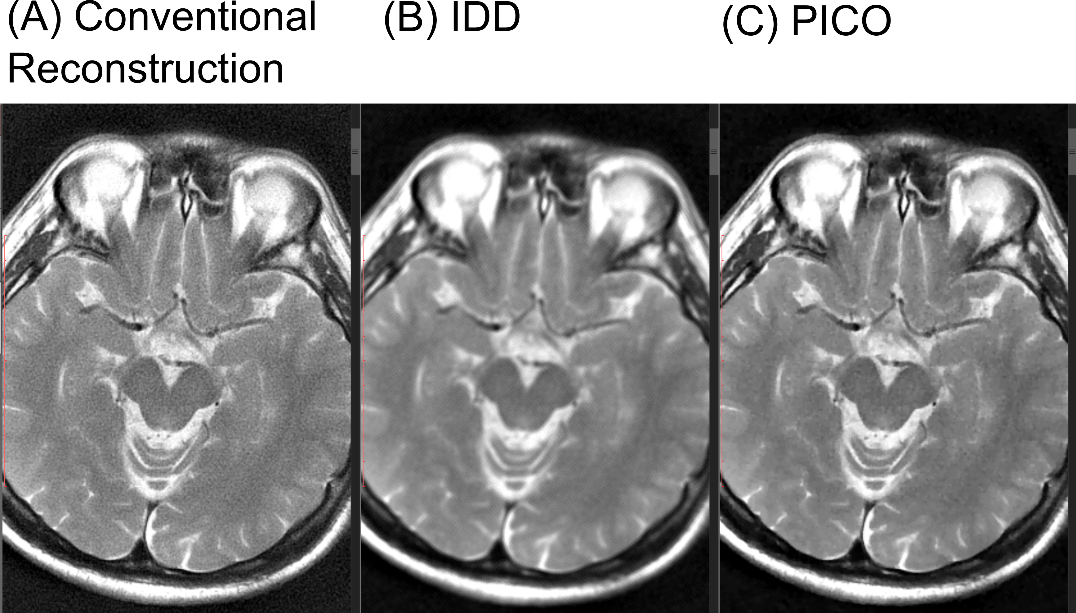
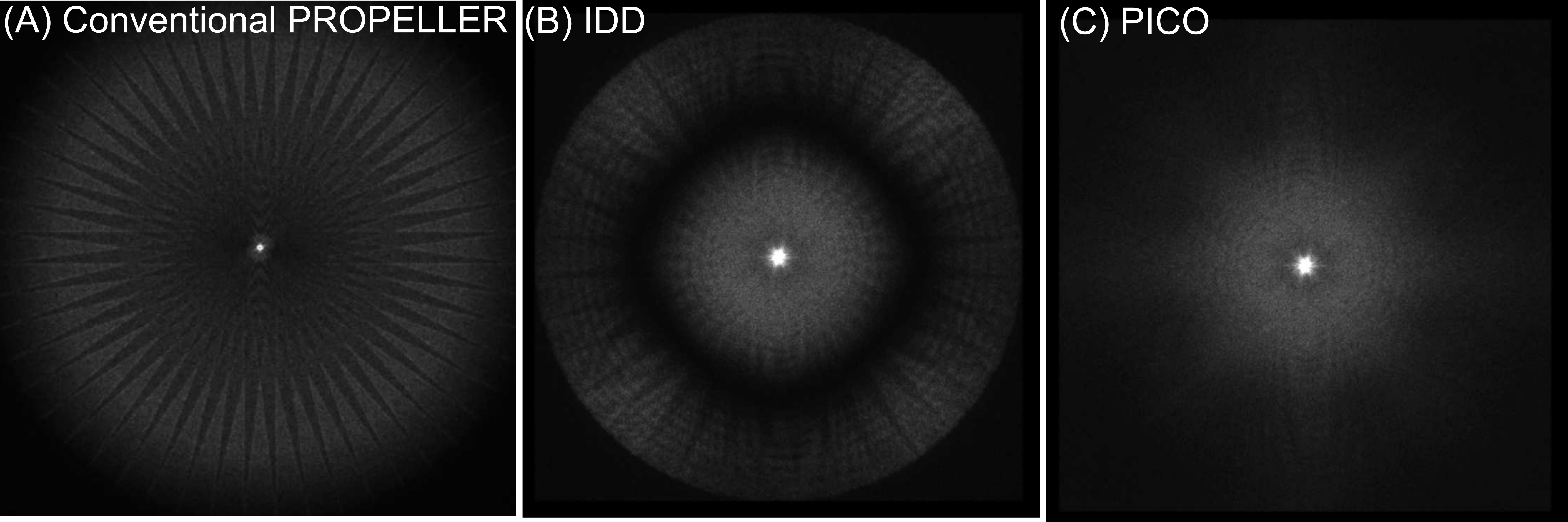
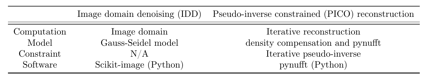
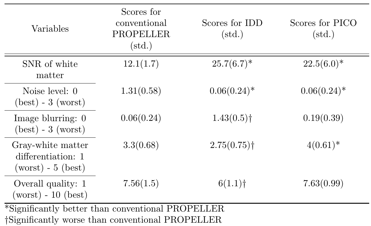
Table 2: Subjective quality assessment by radiologists.
*Significantly better than conventional PROPELLER
†Significantly worse than conventional PROPELLER