1513
Reducing acquisition time while maintaining spatial resolution with extended readouts and R2* modeling1Bioengineering, University of Illinois at Urbana-Champaign, Urbana, IL, United States, 2Beckman Institute, University of Illinois at Urbana-Champaign, Urbana, IL, United States
Synopsis
Single shot readouts are limited in length by field inhomogeneity and R2* relaxation. With the inclusion of a complex field map, existing field corrected reconstruction algorithms can compensate for both field inhomogeneity and R2* relaxation during extended readouts. Results for spiral acquisitions of up to 56ms in length are demonstrated on a human volunteer.
Introduction
The spatial resolution of single-shot imaging has increased as gradient performance and shimming has improved on modern MRI scanners. Ultimately the duration of the data acquisition is limited by magnetic field inhomogeneity, which causes image distortions that increase with readout duration, and R2* based filtering in k-space, which causes a filtering effect at the k-space points acquired at later times. Through the use of model based reconstruction this work demonstrates the use of combined field inhomogeneity and R2* corrections to enable high resolution spiral imaging with fewer shots and longer readouts.Methods
Field inhomogeneity corrections for use with non-Cartesian imaging have been successfully applied using a time segmentation algorithm [1] and iterative reconstruction [2]. This approach primarily addresses one of the limitations of long readouts, namely background field inhomogeneity; however, using this model-based approach, R2* relaxation can be included in the signal model correcting the associated filtering effect in k-space. With both R2* relaxation and field inhomogeneity modeled, the effects are compensated during the readout to maintain high spatial resolution information in the estimated image. The signal model is easily accommodated in standard field correction algorithms through the inclusion of a complex field map, $$$FM = \omega_e -jR_2^*$$$, and the use of a Hanning temporal interpolator, which captures the time-dependent behavior of the signal during the readout due to the magnetic field map, $$$\omega_e$$$, and the R2* map [1,3-4]. Using this approach, we acquire readouts that are longer (around 56 ms) than typically collected in single-shot imaging (20-30 ms). To illustrate the method, spin echo spiral out acquisitions with 8, 4, or 2 shots were designed with matrix size 192, 240mm FOV, FA 90, TE 16ms, TR 2s, and readout lengths 14ms, 28ms, or 56ms respectively. A sampling point spread function (PSF) was simulated to demonstrate the loss of resolution that can occur from T2* decay during the longest proposed readout of 56ms in length. Data was acquired with a Siemens Magnetom Prisma on a human volunteer. T2* and field maps were jointly estimated for the acquired data using an iterative model [3] and a multi-echo asymmetric spin echo sequence with a spin echo time of 16ms and asymmetric delays of 0, 0.5ms, 1ms, 5ms, 15ms, and 30ms.Results:
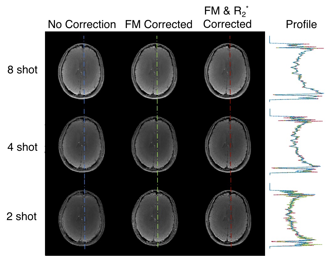
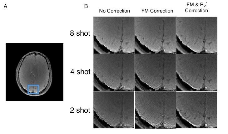
Simulation of sampling PSF with and without the effect of R2* decay showed a loss of effective resolution of approximately 20% for an average T2* value of 42.6ms as measured from the R2* map. For a 192 matrix acquisition, T2* in a two shot acquisition would limit the information in the image effectively to 164 matrix size without correction. Figure 1 shows the results of the proposed acquisition with and without the R2* corrections included, as well as a line plot comparing the reconstructions. Figure 2 shows an inset view (A) with the acquisitions and corrected reconstructions methods. R2* relaxation incorporated into the reconstruction lead to improved spatial resolution, even at the longest two shot readout as shown in the inset.Discussion and Conclusions:
This abstract addresses one of the major factors limiting the lengths of readouts in MRI and hence the spatial resolution of single-shot acquisitions. Long readout single-shot acquisitions are desirable when used in combination with signal mechanisms that require time to develop contrast, such as in BOLD functional MRI and in diffusion weighted imaging. The proposed correction is compatible with conventional parallel imaging techniques such as SENSE [5]. As an example, the two shot acquisition could be made into a single shot acquisition with only an easily achieved parallel imaging reduction factor of 2, whereas the 8 shot data would require a challenging reduction factor of 8 to achieve the same total acquisition time. The proposed approach will enable significant increases in spatial resolution of acquisitions without significant increases in acquisition times.Acknowledgements
This work was supported by NIH F30AG055283 and R01EB018107.References
[1] Sutton BP, Noll DC, Fessler JA. Fast, iterative image reconstruction for MRI in the presence of field inhomogeneities. IEEE Transactions on Medical Imaging. 2003;22(2):178–188.
[2] Fessler JA. Image Reconstruction Toolbox. Available from: https://web.eecs.umich.edu/~fessler/code/
[3] Ngo GC, Sutton BP. R2* Mapping for Robust Brain Function Detection in the Presence of Magnetic Field Inhomogeneity, Proceedings IEEE EMBC Chicago, pp.1537,1540, 26-30 August 2014.
[4] Olafsson VT, Noll DC, Fessler JA. Fast Join Reconstruction of Dynamic R2* and Field Maps in Functional MRI. IEEE Transactions on Medical Imaging. 2008.
[5] K. P. Pruessmann, M. Weiger, M. B. Scheidegger, and P. Boesiger, “SENSE: Sensitivity encoding for fast MRI,” Magnetic Resonance in Medicine, vol. 42, no. 5, pp. 952–962, Nov. 1999.
Figures