1511
Assessing intrinsic velum height in vowels using time-resolved MRI1Medical Physics Group, Institute of Diagnostic and Interventional Radiology, Jena University Hospital - Friedrich Schiller University Jena, Jena, Germany, 2Institut für Germanistische Sprachwissenschaft, Friedrich Schiller University Jena, Jena, Germany, 3Michael Stifel Center for Data-driven and Simulation Science Jena, Friedrich Schiller University Jena, Jena, Germany, 4Abbe School of Photonics, Friedrich Schiller University Jena, Jena, Germany, 5Center of Medical Optics and Photonics, Friedrich Schiller University Jena, Jena, Germany
Synopsis
Real-time MRI and synchronised audio were used to examine intrinsic velum height in German vowels. Two adult female subjects produced five repetitions of a set of sentences containing, among other target material, the point vowels /i: a: u:/ in the same phonologically non-nasalised context. Even in this small sample, the subjects exhibit variation in velum height across vowel categories, and show considerable interindividual variation in velum height during the production of the same vowel category. Collection of further data from normal subjects will be used to create a robust baseline for the assessment of abnormal velum activity.
Purpose
Measuring the auditory, acoustic and articulatory correlates of nasality is notoriously difficult1. Many methods of measuring nasality are relatively indirect and difficult to interpret, e.g. nasalance2. More direct methods of measuring velum height, such as velotrace3, which uses a lever sitting directly on the upper surface of the velum is invasive and very uncomfortable for the subject. Midsagittal time-resolved MRI offers a non-invasive way of observing velum height (and shape). In recent years continuing improvements in the temporal resolution of MRI4,5 have allowed us to examine dynamic velum activity in running speech both intrinsic to different sounds, as well as in coarticulatory patterns6. The present study uses time-resolved MRI and synchronised audio to record speakers producing vowels in the same phonologically non-nasal sentence contexts to examine intra- and interindividual variation in intrinsic velum activity.Material and Methods
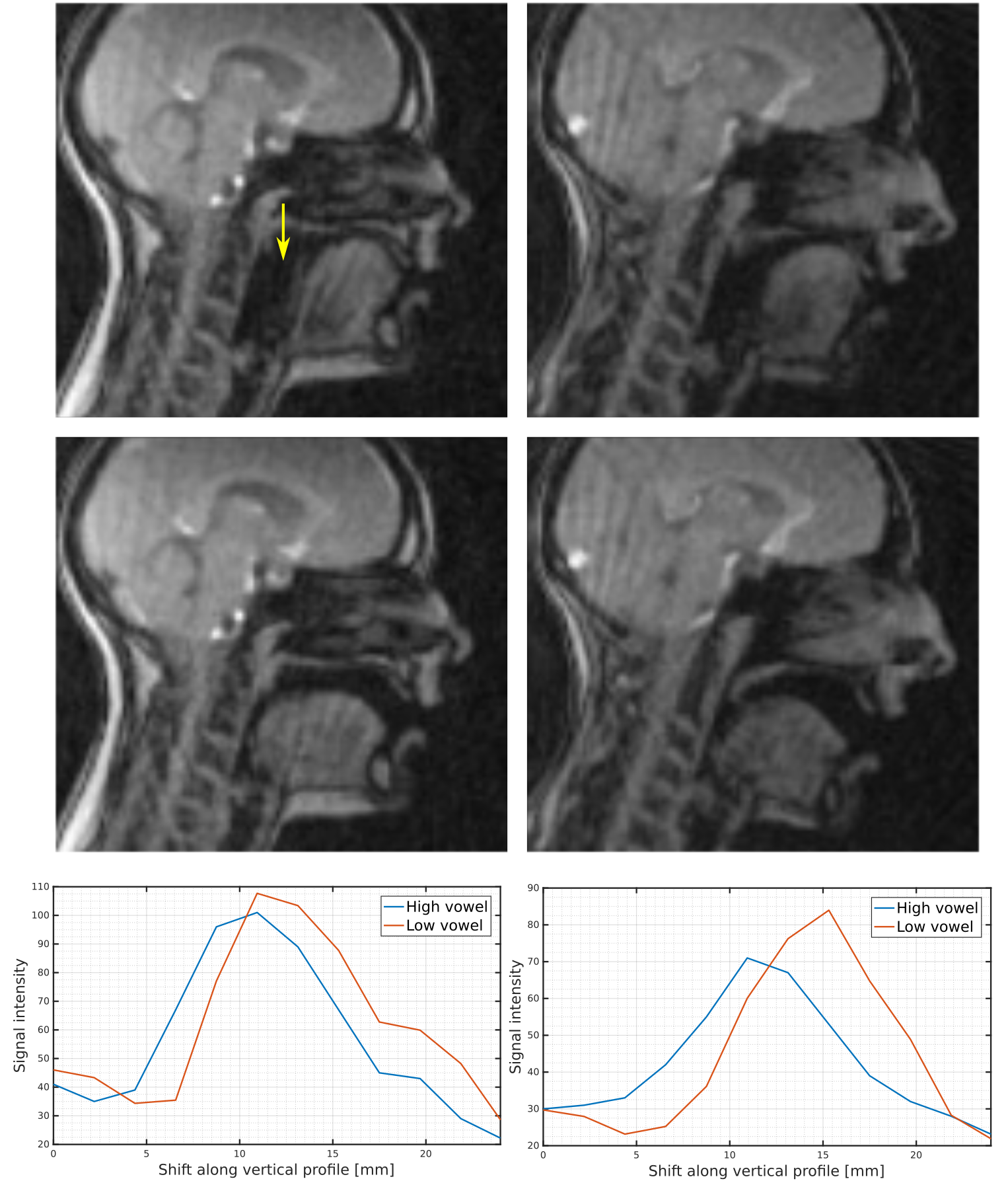
For image acquisition an undersampled radial real-time imaging sequence using rotation angles based on the golden-ratio7 was used with parameters: 128 x 128 acquisition matrix size, 88 radial readouts per frame, 3.2 ms TR, 1.3 ms TE, 280 x 280 mm² FOV, 7 mm slice thickness and 15° flip angle. Measurements were performed on a clinical 3 T Siemens Prisma fit scanner using a vendor supplied 20 channel head coil. Two female German speakers were recorded producing 27 randomly ordered German sentences five times. Acoustic recordings were made using one optical microphone (MO 2000, Sennheiser) placed in 3 cm distance to the subject's mouth. Image reconstruction was performed offline using MATLAB, 2D regridding with iterative density compensation8 and sliding-window reconstruction9. For image analysis EdgeTrak10 was used to obtain and export contours of the velum during the production of vowel sequences of the German point vowels /i: a: u:/ elicited from abbreviation (AUU, BII, IAA) embedded in regular sentences, e.g. Sie fuhren letzte Woche zur BII ganz schnell (“Last week they drove to the BII very fast”), as well as the vowels /a: u:/ in the context of proper nouns (Gabi, Gubi).Results
As expected from earlier studies, even in the phonologically non-nasal context, high vowels /i:/ and /u:/ are accompanied by higher positions of the velum than the low vowel /a:/. While subject FN only exhibits a slight lowering of the velum going from /i:/ or /u:/ to /a:/, subject SN exhibits a more marked lowering of the velum showing a clear opening of the velopharyngeal port. Figure 1 shows frames from the articulation of the high vowel /i:/ (top) and the low vowel /a:/ (bottom) for subjects FN (left) and SN (right) with profile lines drawn through the velum. Dynamic information reveals that in a vowel sequence, such as /i: a: a:/ (IAA), velum lowering and tongue lowering seem to be occurring synchronously. The five repetitions of the same vowel sequences show these patterns of velum height to be consistent across both speakers.Discussion and Conclusion
Recent advances in real-time MRI have proven to be a valuable tool for a direct measurement of nasality in an non-invasive and more comfortable way than previously possible with methods like velotrace or nasalance. The present study demonstrated that by using MRI dynamic velum parameters can be reliably estimated even at single-subject level and for multiple repetitions of the same vowels. For future studies a larger sample of normal male and female subjects is to be collected in order to quantify the normal range of velum activity during vowels as a basis for assessing abnormal velum activity in speakers with auditorily abnormal levels of nasality (hypo-/hypernasality).Acknowledgements
No acknowledgement found.References
1. Simpson, AP. The first and second harmonics should not be used to measure breathiness in male and female voices. J Phonetics. 2012;40:477–490.2.
2. Dalston RM, Warren DW, Dalston ET. A preliminary investigation concerning the use of nasometry in identifying patients with hyponasality and/or nasal airway impairment. J Speech Hear Res. 1991;34(1):11–18.3.
3. Horiguchi, S, Bell-Berti, F. The Velotrace: A device for monitoring velar position. Cleft Palate J. 1987;24(2):104–111.4.
4. Uecker M, Zhang S, Voit D, Karaus A, Merboldt KD, Frahm J. Real-time MRI at a resolution of 20 ms. Magn Reson Med. NMR Biomed. 2016;23(8):986–94.
5. Niebergall A, Zhang S, Kunay E, Keydana G, Job M, Uecker M, Frahm J. Real-time MRI of speaking at a resolution of 33 ms: undersampled radial FLASH with nonlinear inverse reconstruction. Magn Reson Med. 2013;69(2):477–85.
6. Blaylock, R, Louis G, & Shrikanth N. Velum control for oral sounds. In Proc. Interspeech. 2016;1084–1088.
7. Winkelmann S, Schaeffter T, Koehler T, Eggers H, Doessel O. An optimal radial profile order based on the Golden Ratio for time-resolved MRI. IEEE Trans Med Imaging. 2007;26(1):68–76.
8. Zwart NR, Johnson KO, Pipe JG. Efficient sample density estimation by combining gridding and an optimized kernel. Magn Reson Med. 2012;67:701–710.
9. Riederer SJ, Tasciyan T, Farzaneh F, Lee JN, Wright RC, Herfkens RJ. MR fluoroscopy: Technical feasibility. Magn Reson Med. 1988;8:1–1510. Li, M, Kambhamettu, C, Stone, M. Automatic contour tracking in ultrasound images. Clin Linguist Phonet. 2005;19(6–7):545–554.
Figures