1499
Tri-Fast Spin Echo: A Minimalistic Cross-Platform Multi-Spectral qMRI Pulse Sequence for Routine Clinical UseNing Hua1, Mitchell Horn2, Stephan Anderson3, and Hernan Jara1
1Boston University, Boston, MA, United States, 2Boston University, MA, United States, 3Boston University
Synopsis
Purpose: To develop a simple and cross-platform pulse sequence that meets the following criteria: 1) all directly acquired images are clinically useful, 2) achieves minimalistic scan times, and 3) leads to excellent image quality as well as accurate MS-qMRI mapping. Methods: Tri-FSE consists of a single-echo-FSE sequence that is run in temporal concatenation with a dual-echo FSE sequence that all together generate T1-, T2, and PD-weighted directly acquired images. Results: Tri-FSE was implemented at medium and high spatial resolution at 1.5T and 3.0T. Conclusion: Tri-FSE is a dual purpose simple pulse sequence that is useful for clinical and scientific purposes.
Introduction
The directly acquired images generated with multi-spectral (MS) qMRI --proton density (PD) and relaxation times (T1, and T2) -- pulse sequences generally do not conform to the radiologist’s contrast expectations and consequently MS-qMRI is largely relegated to research endeavors. Because scientific research can reduce patient throughput there is great need for developing dual-purpose MS-qMRI pulse sequences that can simultaneously be used as clinical as well as scientific tools. The purpose of this work was to develop and implement a minimalistic and cross-platform pulse sequence --termed herein Tri-FSE-- that meets the following criteria: 1) any and all directly acquired images are clinically useful, 2) achieves minimalistic scan times, and 3) leads to excellent directly acquired image quality as well as accurate MS-qMRI mapping.Methods and Materials
As shown in Figure 1, Tri-FSE consists of a single echo-FSE sequence that is run in temporal concatenation --and with the same pre-scan settings-- with a dual-echo FSE sequence that all together generate T1-, T2, and PD-weighted directly acquired images. Tri-FSE was implemented at standard clinical spatial resolution (total combined scan time ≤ 7min. voxel dimensions = 0.5 x 0.5 x 5mm3) at 1.5T (Achieva, Philips Healthcare, Best, The Netherlands) and 3.0T (Discovery MR750w, GE Healthcare, Waukesha, WI). Key parameters: 22/100ms effective echo times, 600/6,000ms repetition times (values depending on anatomical coverage). Tri-FSE was also implemented at high spatial resolution (3T: total scantime 10min, 90 slices with reconstructed voxel 0.47 x 0.47 x 1.8mm3). In all cases, the echo train length of the DE-FSE (ETL=10-14) was twice that of the single echo T1-weighted sequence (ETL=5-7), parallel imaging was not used, and scanning was performed with the quadrature body coil and a multi-channel phased array head coil for RF transmission and signal detection respectively. The matching PD, T1, and T2 qMRI mapping algorithms were programmed in Mathcad (version 2001i, PTC, Needham MA) and adapted to handle large datasets. Tri-FSE was implemented clinically at 1.5T and 3.0T and in addition, one healthy volunteer (male, 24yo) was consented and scanned at 3T with the high-spatial resolution variant of the Tri-FSE in accordance and approval by our Institutional Review Board (IRB).Results
The Tri-FSE pulse sequence was implemented in the scanners of our institution and is run routinely for clinical purposes. An example of the high spatial resolution Tri-FSE directly acquired images is illustrated in Figure 2 (top row). The directly acquired images conform to clinical standards in terms of PD-, T1-, and T2- weighting and meet or exceed current spatial resolution standards. The corresponding qPD, qT1, and qT2 maps are shown in Figure 2 (bottom row): ROI measurements for white matter (WM), cortical gray matter (GM), intraventricular cerebrospinal fluid (CSF) are approximately WM (0.5ru/600ms/90ms), GM (0.8ru/1,400ms/110ms), and CSF (0.96ru/3,100ms/2,300ms).The values for GM and CSF are in the range of reported 3T values (1) at age 24 years. Notably, the T1 values obtained for WM appear underestimated (see Discussion).Discussion
With the advent of faster MRI pulse sequences and image processing techniques, MS-qMRI is now possible in routine clinical practice. Availing MS-qMRI datasets of the main parameters enable several post-processing applications such as Synthetic MRI, Structural qMRI, and most importantly, it can enhance diagnostic accuracy by allowing direct intra-patient and inter-patient comparisons. Tri-FSE described herein is scan time efficient and can be used with scanners any manufacturer that offer the DE/SE-FSE pulse sequences; it generates directly acquired images that are immediately available for clinical assessment and further offer the potential for MS-qMRI processing. The T1 underestimation for WM likely results from the strong magnetization transfer effects that are inherent to the RF-intensive Tri-FSE pulse sequence (2, 3).Conclusion
Tri-FSE has been implemented and tested at 1.5T and 3.0T: it is a scan time efficient and widely available pulse sequence for MS-qMRI of PD, T1, and T2. This simple and minimalistic approach to MS-qMRI could lead to broadening the clinical use of qMRI measures for improved assessment of disease as well as enrich scientific research. Further theoretical work is necessary for correcting the inherent MT effects.Acknowledgements
No acknowledgement found.References
1. Stanisz GJ, Odrobina EE, Pun J, et al. T1, T2 relaxation and magnetization transfer in tissue at 3T. Magn Reson Med 2005;54(3):507-512. 2. Melhem ER, Jara H, Yucel EK. Multislice T1-weighted hybrid RARE in CNS imaging: assessment of magnetization transfer effects and artifacts. J Magn Reson Imaging 1996;6(6):903-908. 3. Meara SJP, Barker GJ. Impact of incidental magnetization transfer effects on inversion recovery sequences that use a fast spin echo readout. Magnetic Resonance in Medicine 2007;58(4):825-829.Figures
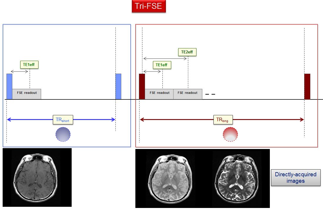
Figure 1: Tri-FSE
consists of a single echo-FSE sequence that is run in temporal concatenation
--and with the same pre-scan settings-- with a dual-echo FSE sequence that all
together generate T1-, T2, and PD-weighted directly acquired images.
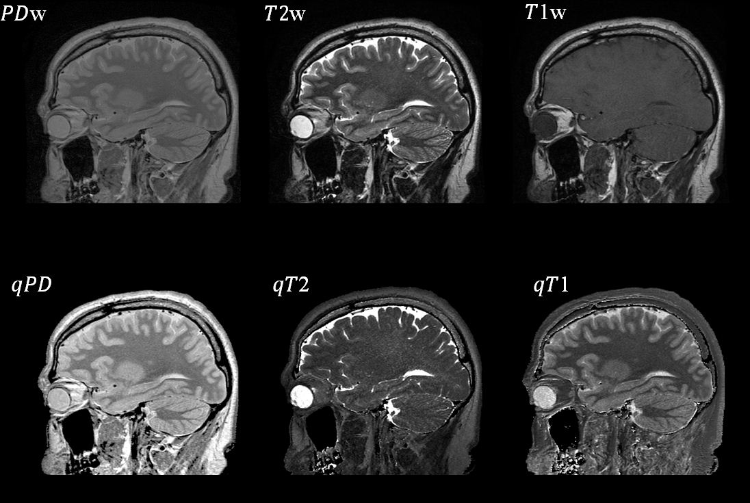
Figure
2: Selected directly
acquired and MS-qMRI maps obtained with the high spatial resolution Tri-FSE
pulse sequence at 3.0T.