1496
Improving Arterial Spin Labeling Acquisition to Reduce the Effect of Delayed Arrival Time1Physics Department, University of Sao Paulo, Ribeirao Preto, Brazil, 2Internal medicine, Medical School of Ribeirao Preto, Ribeirao Preto, Brazil, 3Physics Institute of Sao Carlos, University of Sao Paulo, Sao Carlos, Brazil
Synopsis
Arterial Spin Labeling (ASL) is a powerful technique to evaluate cerebral blood flow. To analyze hemodynamics effects with ASL, multiples acquisitions over the time are realized, which is called multiphase ASL. In conventional multiphase ASL methods, the later phases has low contrast to noise ratio, so it becomes difficult to analyze it. This study purposes a solution to this problem, through a modulation in the acquisition flip angle. With this technique, the flip angle of all phases follows a modulation equation, so that the ASL signal over the phases becomes nearly constant.
Purpose
Arterial Spin Labeling (ASL)1, 2 is a non-invasive method to measure perfusion quantitatively. A radiofrequency pulse labels arterial blood protons, which flow through the vascular tree and exchange water with the unlabeled brain tissue, where MR images are acquired. Usually, a single delay time (TI) between labeling and image acquisition is used. However, since arrival time varies from different brain regions, defining a single TI may compromise proper CBF quantification. Multiphase ASL was proposed to overcome that issue. In this case, a set of images are acquired after different TIs. In conventional multiphase ASL acquisition, the contrast-to-noise ratio (CNR) decreases over the phases due the effects of longitudinal relaxation time (T1) and multiple excitations in the image region, the contrast-to-noise ratio (CNR) decreases over the phases, so that analyzes the latter phases becomes difficult. It is especially critical to evaluate patients with cerebrovascular diseases associated with delayed arterial arrival time, such as stroke and carotid stenosis. To overcome this problem, we propose an optimization of the acquisition scheme using variable flip angle.Methods
We optimized the acquisition scheme using variable flip angles determined by setting the condition that the difference between control and labeled images were constant for all phases. In that case, flip angle for the i-th phase is given by3:
$$FA_{i}=\arctan(\frac{\sin(FA_{max})\cdot\exp(-\frac{(m-i)\tau}{T_{1}})}{\sqrt{1+\sum_{k=1}^{m-i-1}\sin^{2}(FA_{max})\cdot\exp(-\frac{2k\tau}{T_{1}})}})$$
Simulation was performed using Matlab to investigate the efficiency of proposed method when compared to the constant flip angle implementation. Healthy adult volunteers (N=20) and patients with severe unilateral carotid stenosis (N=12) were scanned in a 3T Achieva Philips system equipped with high performance gradient system and a 32-channel head coil. Images were acquired using a GE-EPI sequence with the following parameters: TR/TE=4000/18ms, FOV=240x240mm2, matrix=64x64, slice thickness=6mm and 8 phases acquired with TIs ranging from 675ms to 2600ms and 35 ASL acquisitions.
Results
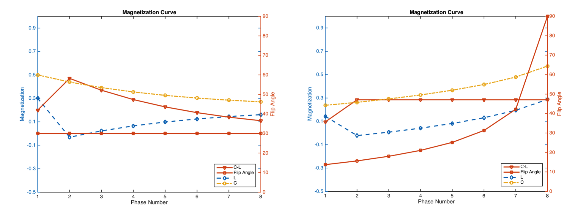
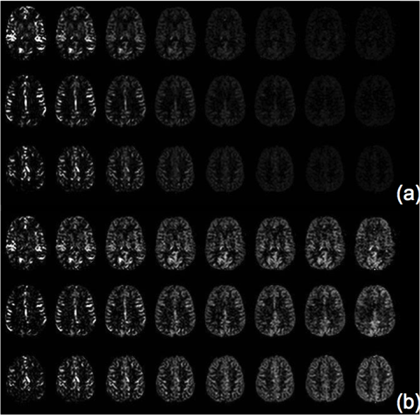
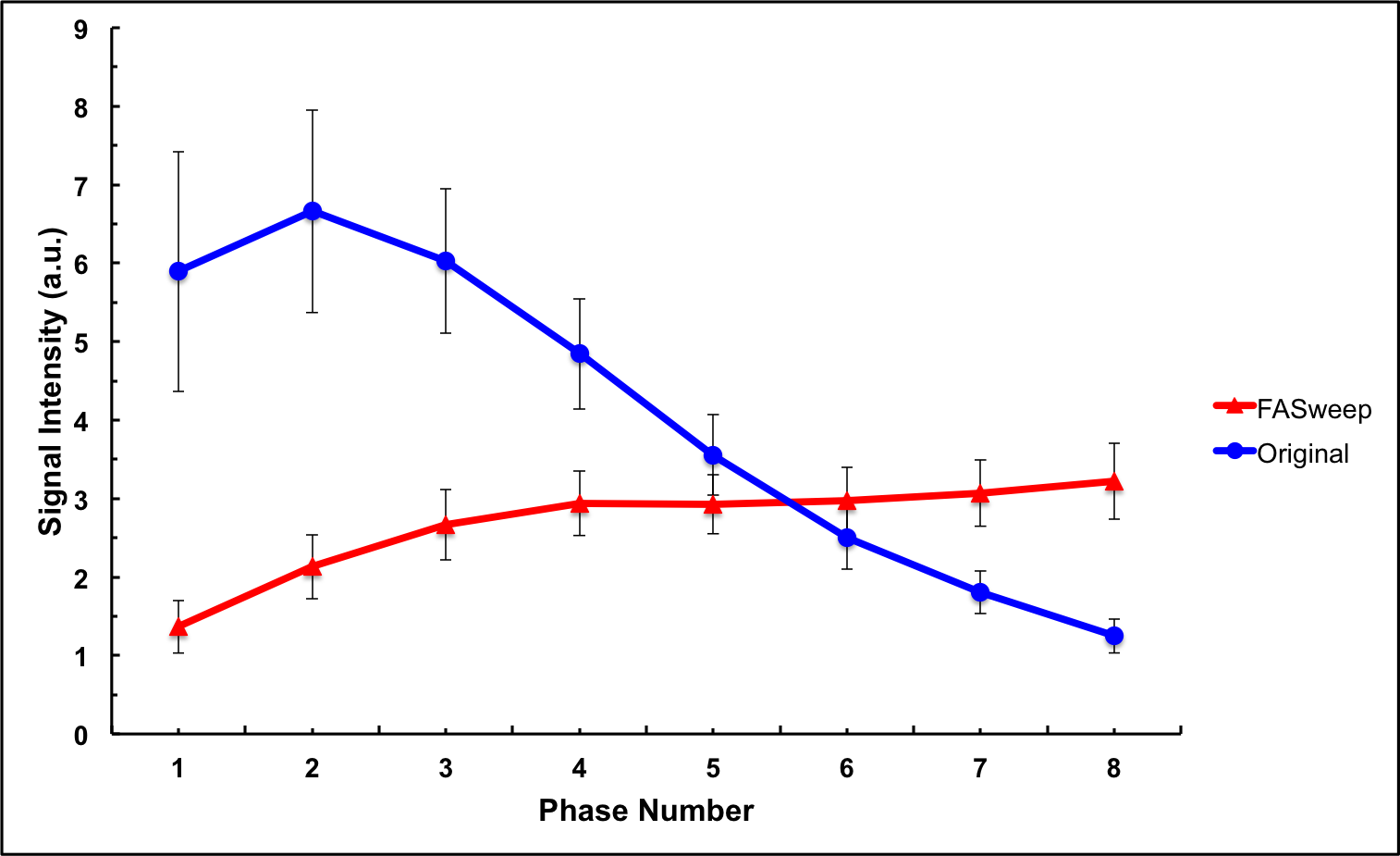
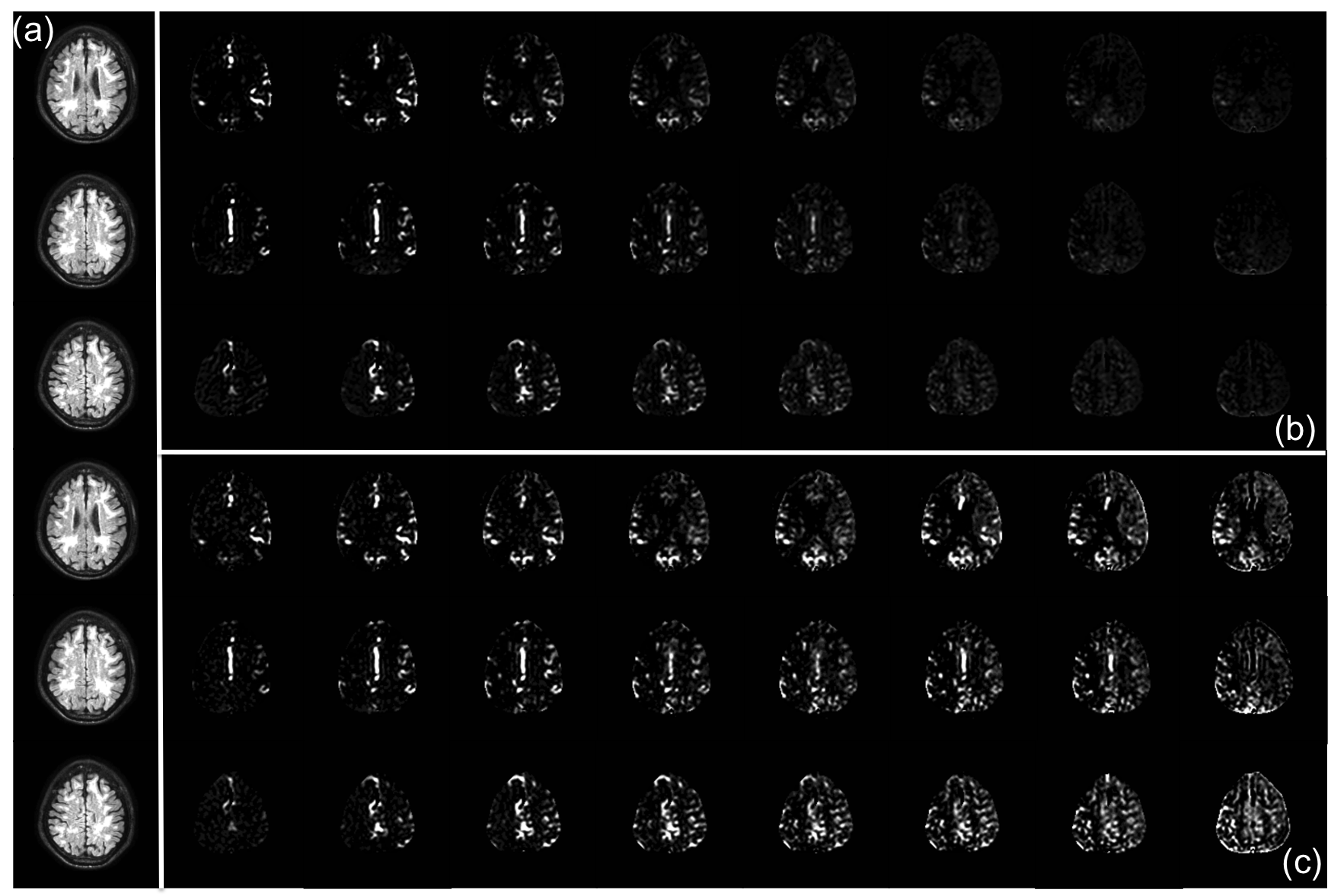
Figure 1 show simulation of the signal intensity for multiphase ASL acquisition using conventional acquisition with constant flip angle (left) and the proposed method (right). In the first case, a systematic reduction of ASL signal can be noticed while modulation of flip angle results in an approximately constant ASL. Figure 2 shows representative multiphase data acquired from a healthy volunteer using conventional approach (a) and flip angle modulation (b). For conventional approach, the average signal intensity shows significant reduction starting from fourth phase while it stays almost unchanged for the proposed method. Figure 3 shows the average ASL signal in gray matter through all phases for healthy volunteers. Figure 4 shows anatomical (a) and multiphase data acquired from a patient with partial occlusion in the right side of internal carotid artery with reduced flow using conventional approach (b) and flip angle modulation (c). The regions irrigated by the right middle and posterior carotid arteries have low perfusion in first phases. Due to low CNR in latter phases, it is difficult to know if blood is arriving while analyzing data obtained using the conventional approach. For the proposed method, it is possible to see that blood starts to perfuse tissue around the 5th phase.Discussion
The results obtained from simulations and healthy controls clearly show that flip angle modulation optimizes the contrast to noise ratio in later phases of multiphase ASL acquisition. Even though according to the simulations the signal intensity of the perfusion-weighted image in a multiple phase acquisition can achieve higher values for earlier phases when using conventional approach, it usually occurs for time points when the spins from labeled blood are in larger vessels and have high signal compromising the accuracy of the CBF estimation. The evaluation of patients with severe unilateral carotid stenosis evidences the importance of the proposed modulation to differentiate regions with delayed transit time from regions with reduced blood flow.Conclusion
The proposed flip angle modulation for multiphase ASL is an efficient way to increase CNR in later phases. This may expand the applicability of the technique, which might become an important tool for helping diagnosing patients with cerebrovascular diseases associated with delayed arterial arrival time.Acknowledgements
CNPq; CAPES; FAPESPReferences
1. Detre, J.A., et al., Perfusion imaging. Magn Reson Med, 1992. 23(1): p. 37-45. 2. Williams, D.S., et al., Magnetic resonance imaging of perfusion using spin inversion of arterial water. Proceedings of the National Academy of Sciences U S A, 1992. 89(1): p. 212-6. 3. Paschoal, A. M.; Leoni, R. F.; Santos, A. ; Foerster, B. U. ; Paiva, F. F. . ASL Contrast Optimization in Multiphase STAR Labeling using Variable Flip Angle. In: Organization for Human Brain Mapping, 2015, Honolulu. Proceedings of the Organization for Human Brain Mapping, 2015. v.
Figures