1494
Ingredients for balanced SSFP Microimaging1Dept. of Radiology, Medical Physics, University Medical Center Freiburg, Freiburg, Germany, 2Bruker Biospin GmbH, Ettlingen, Germany, 3Department of Microsystems Engineering IMTEK, University of Freiburg, Freiburg, Germany, 4Clinics of Radiology and Nuclear Medicine, Radiological Physics, University Hospital Basel, Basel, Switzerland
Synopsis
In this work, balanced SSFP microimaging was succesfully performed in 3D (voxel size 40μm x 40μm x 94μm) and 2D (in-plane resolution 16μm x 16μm) on a 7T small animal system and a small-bore 7T spectrometer with maximally used gradient amplitude of 1.03 T/m. Key for optimzed image quality was the choice of susceptibility matched phantom materials and the proper choice of bSSFP flip angle under consideration of diffusion attenuation due to imaging gradients.
Purpose
Target of the presented work is to enable and to optimize balanced SSFP microimaging, which means imaging of small (<1 cm) samples at high image resolution (~50 μm). Therefore, minimization of susceptibility effects from materials and optimization of the bSSFP flip angle with respect to intrinsic diffusion damping from strong imaging gradients was performed.Methods
Balanced SSFP is an attractive imaging sequence due to high SNR efficiency, but it is very sensitive to off-resonance frequencies and can easily suffer from banding artefacts. In order to minimize the negative influence on field homogeneity from the susceptibility of, e.g., sample holders or implant materials, we have chosen materials with susceptibility close to water. These were PMMA (polymethyl metaacrylate) and PI (polyimide) with susceptibilities relative to water of -0.023ppm and 0.117ppm, respectively1. From both materials, small phantom plates (PMMA thickness 500μm, PI thickness 150μm) were laser structured with details of down to 100μm size. Phantoms were room temperature oxygen plasma treated for two minutes to improve the water wetting of the phantom surface. Phantoms were finally put into 5mm NMR tubes which were filled with tap water. Relaxation times at 7T were determined spectroscopically to be T1=2.0s and T2=1.9s. With a DW-EPI sequence, the diffusion coefficient was determined to 2.47*10-3 mm2/s.
3D Imaging was performed on a Bruker Biospec 70/20 7T small animal system equipped with a cryogenic 2cm surface quadrature coil and with gradients with maximal amplitude of 676mT/m. Balanced SSFP sequences with different image resolution were applied to verify image quality and to optimize the flip angle under consideration of steady-state diffusion damping from the imaging gradients2. For comparison, a non-RFspoiled unbalanced gradient echo sequence was also applied. Besides, bSSFP imaging was also performed on a 7T vertical bore NMR spectrometer with a 5mm linear volume coil and equipped with a Bruker Micro5 standard bore imaging probe with gradient insert delivering 2.95T/m. For shimming, either a first and second order non-localized FID based shim or a localized field map based method was used.
Results
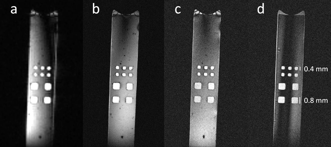
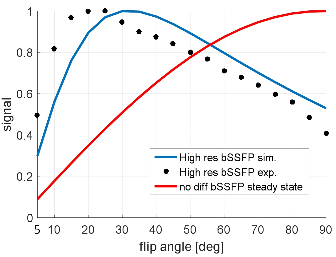
Results of the PMMA phantom imaged with the animal system are displayed in Fig. 1. Banding artefact free images are obtained after field map based shim despite the TR of 8ms. For the low resolution image in Fig. 1a, a series with varying flip angles α shows a maximal signal at approx. 90° according to the normal steady-state formula, while for the high resolution image the maximum signal is obtained at 30° (Fig. 1b) and not at 90° (Fig. 1c). The SNR in dependency of the flip angle is shown in Fig. 2, which compares the experiments of a flip angle series with theory values2 including diffusion effects of the imaging gradients. Note the shift of the experimental curve (obtained after standard SNR analysis) relative to the theoretical values which is probably due to a flip angle miscalibration of 5° of the surface coil signal. Besides, a non-RF-spoiled unbalanced SSFP sequence (spoiled GRE) is shown in Fig 1d. Here, maximal signal is obtained at the Ernst angle of 5°, which shows that diffusion attenuation practically eliminates T2 dependeny of the steady state signal and leads to an SNR that is comparable to the SNR of the 90° bSSFP image in Fig. 1c.
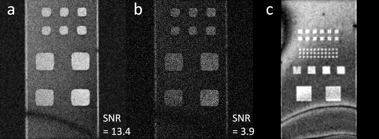
The images obtained with the 2.95T/m gradient in the 7T spectrometer are shown in Fig. 3. A banding artefact is visible here as shimming was only performed with a global first and second order FID-based shim. Maximally used gradient amplitude was 1.03T/m. From the imaging parameters, a SNR increase of factor two would be expected when going from higher (Fig. 3b) to lower (Fig. 3a) resolution. In fact, a factor of 13.4/3.9=3.4 is observed here, which is again explained by the stronger diffusion attenuation2 on the high resolution image. The banding artefact is less prominent in Fig. 3b, as due to strong diffusion damping the bSSFP sequence loses its refocusing behavior and approaches the Ernst regime of a spoiled sequence. Fig. 3c shows the PI phantom with smallest structure size of 100 μm. Imaging of this material with a tolerable banding artefact is feasible.
Conclusion
We have demonstrated the feasibility of bSSFP microimaging up to an in-plane resolution of 16μmx16μm. For microimaging with bSSFP, it is important to take the diffusion attenuation effect of the imaging gradients into account to optimize signal optimization and to make it SNR-superior over spoiled gradient echo imaging. Besides, materials for sample containers, implants or interventional devices have to be carefully chosen, as their susceptibility should match the water susceptibility.
Acknowledgements
This research was supported by the BrainLinks-BrainTools Cluster of Excellence funded by the German Research Foundation (DFG, grant number EXC 1086).References
1. Wapler M, Leupold J, Dragonu I, von Elverfeldt D, Zaitsev M, Wallrabe U. Magnetic properties of materials for MR engineering, micro-MR and beyond. J Magn Reson 2014;242:233-242.
2. Bär S, Weigel M, von Elverfeldt D, Hennig J, Leupold J. Intrinsic diffusion sensitivity of the balanced steady-state free precession (bSSFP) imaging sequence. NMR Biomed 2015;28(11):1383-1392.
Figures