1483
Slice-Accelerated Single-Shot Variable-Flip-Angle Fast Spin Echo with Very Long Echo Trains1Department of Biomedical Engineering, Sungkyunkwan Univiersity, Suwon, Korea, Republic of, 2Center for Neuroscience Imaging Research, Institute for Basic Science (IBS), Suwon, Korea, Republic of
Synopsis
A Fast spin echo (FSE) pulse sequence has been a main workhorse for clinical imaging due to its flexible contrast. Simultaneous multi-slice (SMS) FSE in [1] was shown to be efficient for slice acceleration without much loss of signals. Nevertheless, the previous SMS FSE methods, which employ high-flip-angle, spatially-selective multi-band RF pulses for both excitation and refocusing, still remain sub-optimal due to either high energy deposition or elongated echo spacing (ESP) and thereby limited echo train length (ETL). The purpose of this work is to develop a novel, slice-accelerated single-shot variable-flip-angle (VFA) FSE with very long echo trains, in which multi-band RF pulses are used only for excitation while short hard pulses with VFA are utilized along the refocusing pulse train, enabling very short ESP and very long ETL and thus enhancing imaging efficiency. It is shown that the proposed method makes it possible to complete whole brain imaging within 30 sec without apparent artifacts and noise.
Introduction
A Fast spin echo (FSE) pulse sequence has been a main workhorse for clinical imaging due to its flexible contrast. Simultaneous multi-slice (SMS) FSE in [1] was shown to be efficient for slice acceleration without much loss of signals. Nevertheless, the previous SMS FSE methods, which employ high-flip-angle, spatially-selective multi-band RF pulses for both excitation and refocusing, still remain sub-optimal due to either high energy deposition or elongated echo spacing (ESP) and thereby limited echo train length (ETL). The purpose of this work is to develop a novel, slice-accelerated single-shot variable-flip-angle (VFA) FSE with very long echo trains, in which multi-band RF pulses are used only for excitation while short hard pulses with VFA are utilized along the refocusing pulse train, enabling very short ESP and very long ETL and thus enhancing imaging efficiency. It is shown that the proposed method makes it possible to complete whole brain imaging within 30 sec without apparent artifacts and noise.
Sequence design and Reconstruction
Sequence design: A schematic and timing diagram of the proposed pulse sequence is shown in Fig. 1, consisting of a relatively long multi-band selective excitation RF pulse followed by non-selective, VFA refocusing pulse trains. Fig.2 demonstrates variables flip angles and its corresponding signal evolution of gray matter (GM), in which variable flip angles are calculated using prescribed signal evolution (exponential-flat-exponential). To maintain the shortest possible ESP in the refocusing pulse train while accommodating long multi-band excitation pulse, the first ESP is correspondingly much longer than the regular ESP. To avoid potential loss of signals due to the non-CPMG condition, the flip angle of the first refocusing pulse is set to 180° to position spin population fully on spin echo pathways. Refocusing pulses are 180° phase-cycled to suppress FID artifacts that are pronounced due to multi-band selective excitation and non-selective refocusing. To better condition aliasing separation in the slice direction, CAIPI gradient blips in the corresponding direction are set and rewound on either side of the readout to induce additional spatial modulations.
Reconstruction: To avoid undesirable constant phase accumulations for off-center slices due to the CAIPI gradient blips, phase correction is to be applied. After the phase-cycled averaging over the two acquisitions, SMS reconstruction is performed using the recently proposed SMS-HSL in which a slice-specific null space, which is found using Hankel subspace learning, is employed to suppress slices of no interest while passing only a slice of interest 2.
Materials and Method
Four slices were acquired using the proposed, slice-accelerated single-shot VFA FSE (SMS factor = 2, CAIPI factor = 2) on a 3T whole-body MR scanner. Then, 40 slices of T2-weighted whole brain data were acquired using SMS factors of 5 (CAIPI, 5) and 7 (CAIPI, 5). The imaging parameters were: TR / TE = 3000/417ms (TEeff = 112); resolution=1x1x3mm3; ETL = 256 (single shot); ESP = 3.26ms; bandwidth=781Hz/Pix; and imaging time = 3m, 42s and 30s for single band acquisition, SMS factor = 5, and SMS factor = 7, respectively. Low resolution single-band images were acquired as a reference to construct Hankel subspace.Results
Fig.3 shows the effect of two averages with different phase cycling. As observed in the second row, FID artifacts are substantially pronounced as a results of signal interference such as intensity ripples derived from outer volume slices even though aliasing separation was successfully achieved in SMS-HSL reconstruction. Two acquisition with different phase cycling followed by data averaging eliminates the FID artifacts completely. Figs.4 and 5 represent the reconstructed images and its corresponding intra- and inter-slice leakage maps for five and seven slices of whole-brain data acquired at the SMS factor of 5 and 7, respectively. The SMS-HSL shows robust performance in suppressing image artifacts even though the noise is slightly amplified at the boundaries of brain.
Conclusion
We successfully demonstrated the feasibility of the proposed, slice-accelerated single-shot VFA FSE combined with the SMS-HSL reconstruction, enhancing imaging efficiency without apparent loss of signals and image contrast.Acknowledgements
This work was supported by IBS-R015-D1 and ICT&Future Planning (2016M3C7A1913844).References
1. Borjan A.G, et al. RARE/Turbo Spin Echo Imaging with Simultaneous Multislice Wave-CAIPI. Magn Reson Med. 2015;73:929-938
2. Suhyung P, et al. SMS-HSL: Simultaneous Multi-Slice Aliasing Separation Exploiting Hankel Subspace Learning.Magn Reson Med. 2016|DOI: 10.1002/mrm.26527
Figures
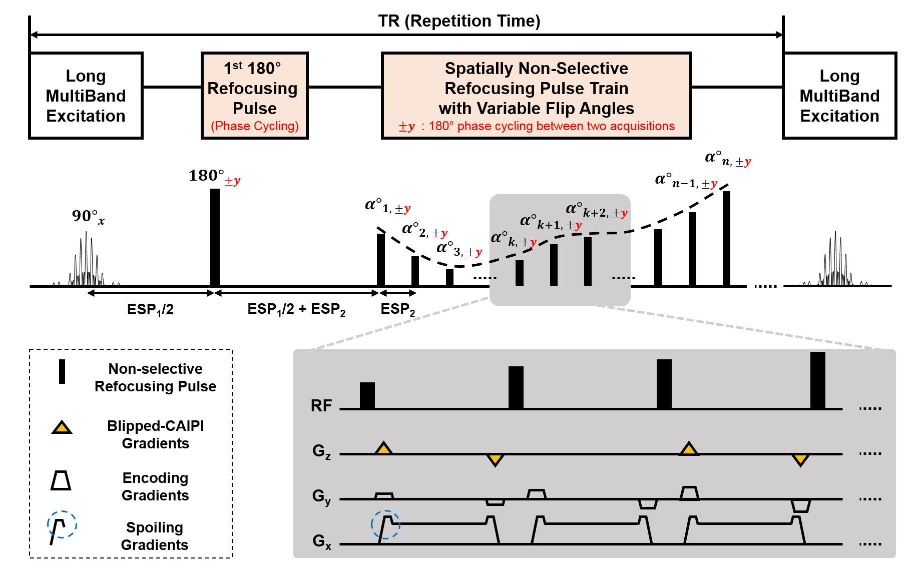

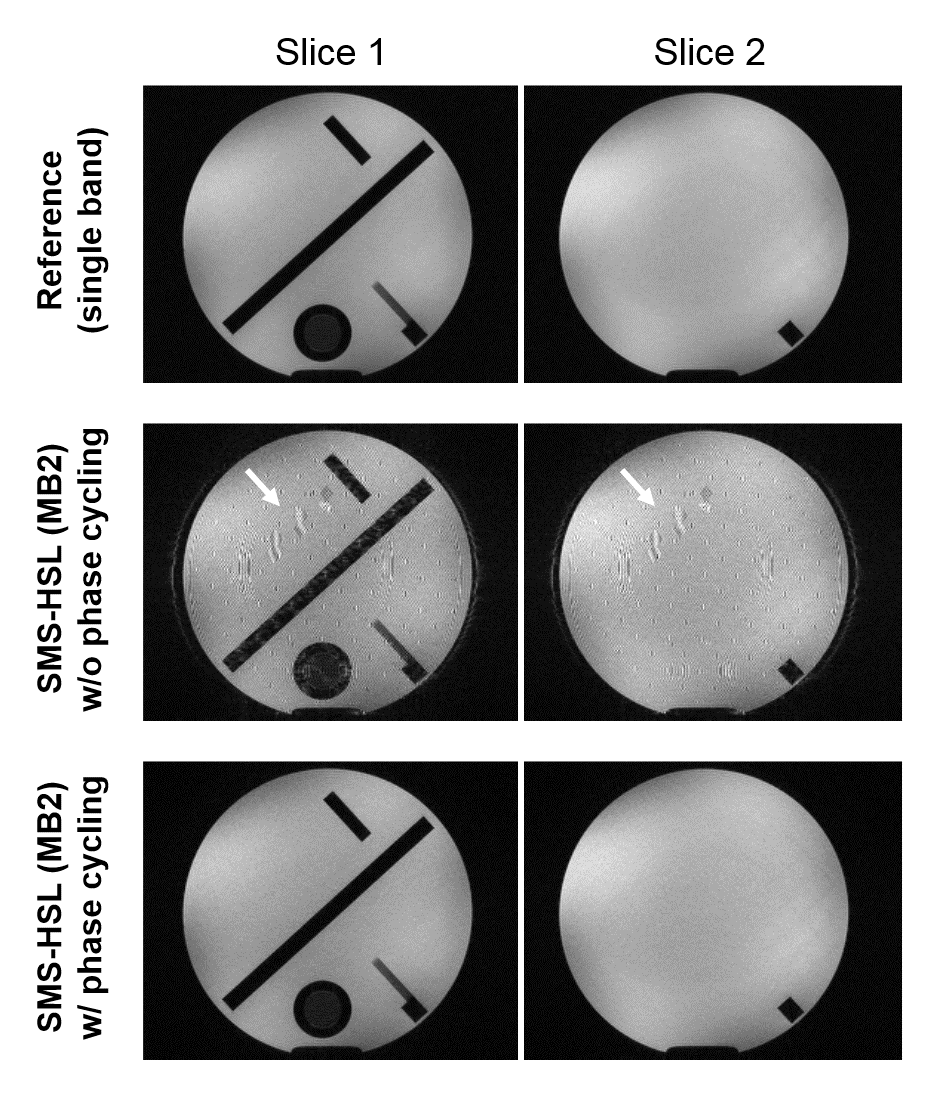
Figure 3. The effect of the phase cycling on FID artifacts. Top row: phantom images obtained from the proposed pulse sequence with phase cycling; Middle and bottom rows: images reconstructed using SMS-HSL acquired using SMS excitation (MB=2) without and with phase cycling, respectively.
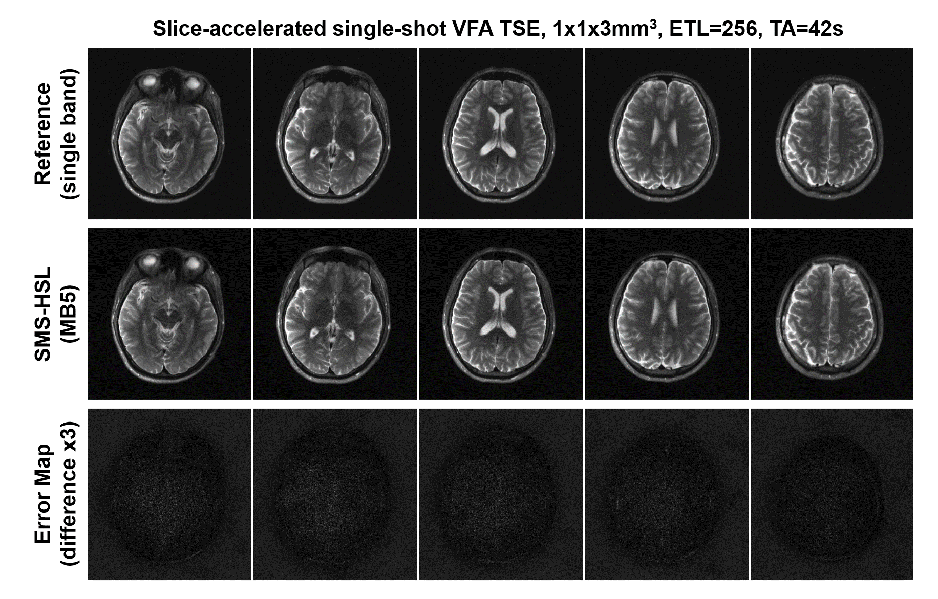
Figure 4. Images and its corresponding error maps reconstructed using SMS-HSL at MB 5. Note that image quality is kept robustly though blurring artifacts slightly appears at the boundaries of brain. Top row: reference images (single band); Middle row: reconstructed images (MB=5); Bottom row: error maps between the reference and reconstructed images.
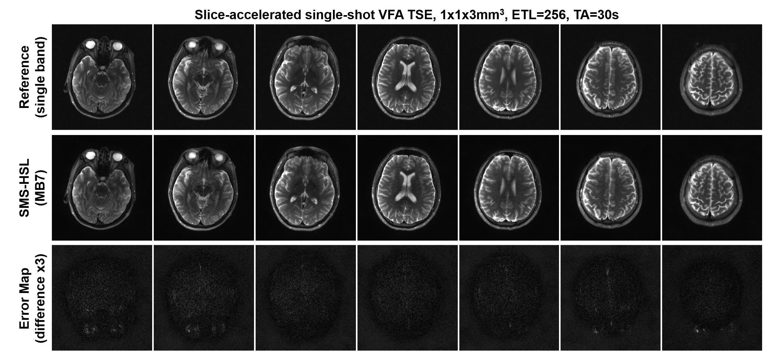
Figure 5. Images and its corresponding error maps reconstructed using SMS-HSL at MB 7. Note that image quality is kept robustly though blurring artifacts slightly appears at the boundaries of brain. Top row: reference images (single band); Middle row: reconstructed images (MB=7); Bottom row: error maps between the reference and reconstructed images.