1464
Bio-inspired optimization of technical fiber-reinforced ramifications using high-resolution MRI of Dracaena marginata branchings as concept generators1Botanical Garden, University Freiburg, Plant Biomechanics Group, Freiburg, Germany, 2Freiburg Centre for Interactive Materials and Bioinspired Technologies (FIT), Freiburg, Germany, 3Freiburg Centre for Interactive Materials and Bioinspired Technologies (FIT), Freiburg, 4Competence Network Biomimetics, Germany, 5Karlsruhe Institute of Technology, Institute of Microstructure Technology, Eggenstein-Leopoldshafen, Germany, 6Department of Radiology University Medical Center Freiburg, Medical Physics, Freiburg, Germany
Synopsis
MRT is still a little-known and highly underestimated imaging method within the field of functional morphology and biomechanics of plants and biomimetics. Its non-invasive and non-destructive character in combination with a large variety of applicable imaging sequences, gives this method a strong potential to shed light to various unanswered scientific questions concerning both the plant structure and function as well as on physiology. Using a Bruker Biospec 94/20 9.4T and a 3D FLASH sequence we could gain new insights into the biomechanics and development of dragon tree ramifications as a source of inspiration for the optimization of technical fiber-reinforced ramifications.
Purpose
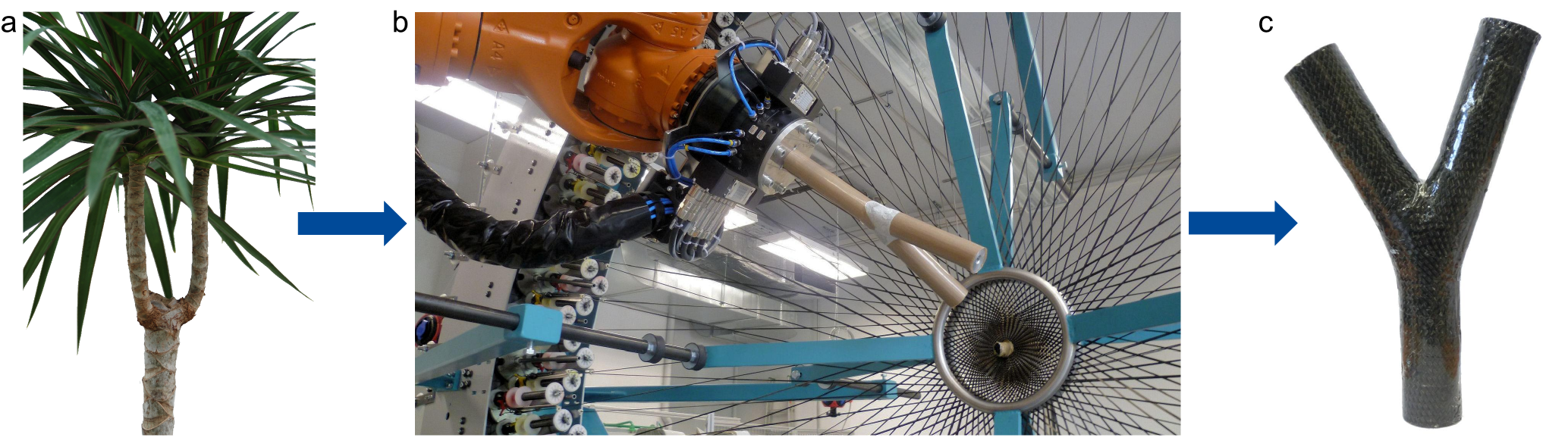
Biomimetics is a scientific field in which nature serves as a source of inspiration for the optimization and/or development of novel technical products1, 2. The aim is to understand the form-structure-function principles of the biological role model to abstract and transfer the findings into technical application2. Common methods, however, are often highly invasive and/or destructive and usually consider only one aspect of interest. Within the field of functional morphology and biomechanics of plants and bimimetics, magnetic resonance imaging has recently proven to be a unique, yet underestimated method enabling novel answers to various scientific questions. In our study, the branch-stem-attachment regions of an arborescent monocot (dragon tree, Dracaena marginata) are used as concept generators for the optimization of technical fiber-reinforced branchings (Fig. 1). With the help of high-resolution magnetic resonance imaging we were able to gain new insights into the functional morphology, biomechanics and development of dragon tree ramifications.
Methods
T2-weighted images were acquired on a Bruker Biospec 94/20 9.4 T using a 3D FLASH sequence (voxel: 100 µm isotropic, acquisition time: 13 h) and quadrature volume coil with 7 cm inner diameter. Image post-processing was conducted with “3D-Slicer”3 and Fiji4. In order to identify a load-adapted placement of mechanically relevant tissues, the branch-stem-attachments of dragon trees (Dracaena marginata) were repetitively imaged in an unloaded and subsequently a mechanically loaded (bending force: 150N) condition. Additionally, the branch ontogeny was analyzed in a second set of experiments by decapitating a plant and repetitively imaging the induced development of 4 buds in the course of a long-term study (180 days). The apical stem segment was imaged every 5 days until day 50 and in the following every 30 days until day 180.Results
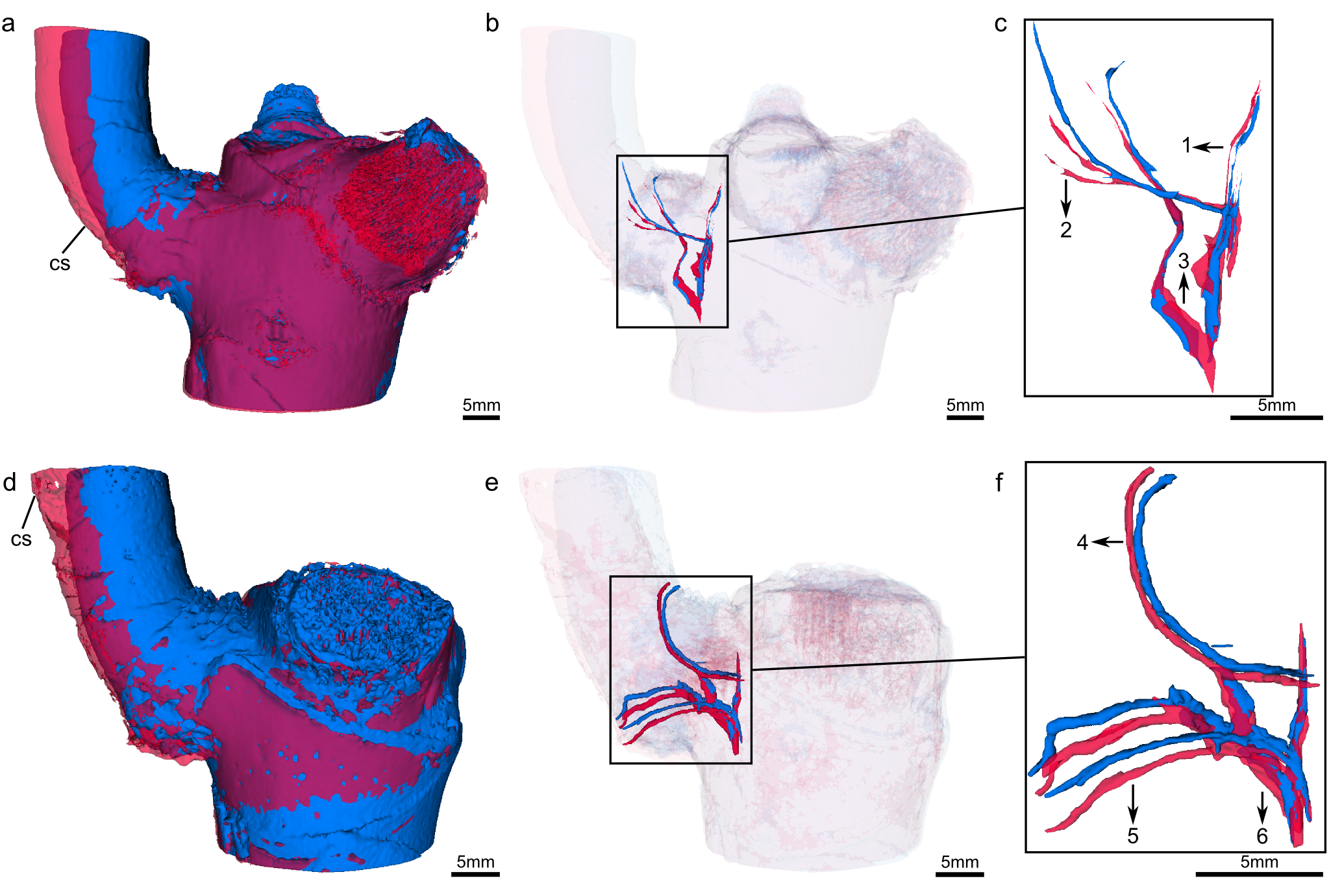
The biomechanical experiments revealed a load-adapted arrangement of mechanically relevant tissues (the vascular bundles and their fiber caps) within the branch-stem-attachment region5 (Fig 2). The vascular bundles are always aligned in direction of occurring stress trajectories. Depending on the orientation within the stem-branch-attachment region they either have to withstand tensile loads or are pressed into the soft matrix of parenchyma tissues thus cushioning compressive loads.
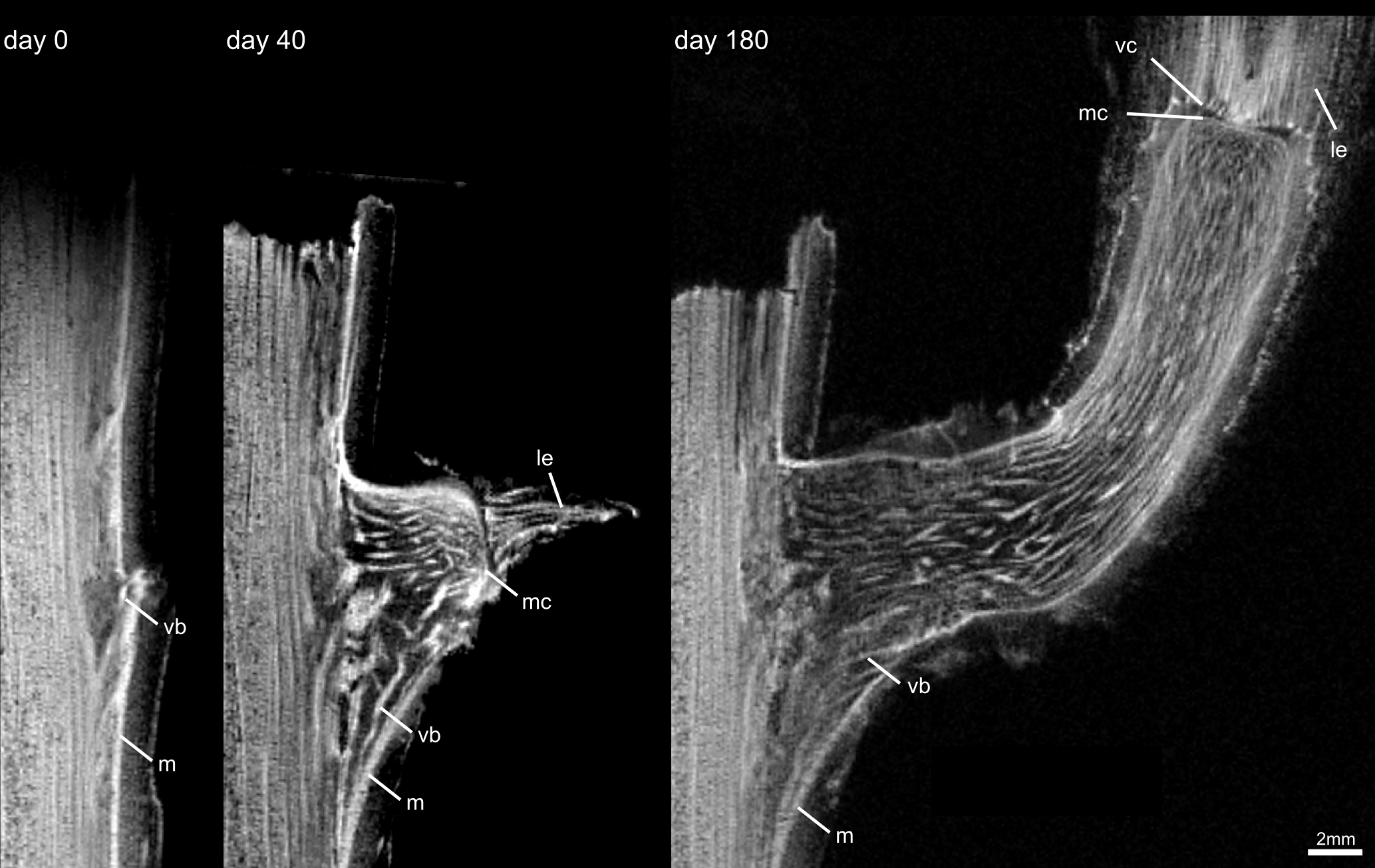
In the course of the long-term study analyzing the ontogeny of the branch-stem-attachment region it became possible to 3D image the growth of 4 buds. One bud decayed within the first 25 days (b1, closest to the decapitation region) and the bud furthest away from the decapitation region (b4) paused growth completely after 20 days. Bud b2 and b3 differed in their rate of growth, with bud 2 having the highest level of development (see ontogeny of bud 2 in Fig. 3). A comparative study of all buds made it possible to identify distinct ontogenetic stages. They help to clarify the successive development of the load-adapted arrangement of the vascular bundles.
Discussion
The identification of a load-adapted arrangement of mechanical relevant tissues and the analysis of the ontogenetic development within the branch-stem-attachment regions of Dracaena marginata will be transferred into novel improved technical fiber-reinforced ramifications. This can be achieved by a dual optimization: The fabrication process can be optimized according to the findings of the results of the analysis of the ontogenetic development. Consequently, the technical fiber-reinforced ramification will be optimized as the technical fibers (carbon, aramid etc.) are placed in a manner comparable to the biological role model.Conclusion
In the field of functional morphology and biomechanics of plants and biomimetics the potential of MRI as a non-destructive and non-invasive imaging method has, until now, been underestimated. The combination of anatomical, biomechanical and developmental studies enabled us to analyze both the growth and functional morphology of dragon tree branch-stem-attachments helping to understand the biomechanical importance of the structures. This methodical approach does mot only generate new knowledge in the field of plant science, but also enhances the knowledge transfer between different disciplines of our interdisciplinary biomimetic project, resulting in novel technological products.Acknowledgements
The authors LH, TM and TS thank the DFG Priority Program SPP 1420 ‘Biomimetic. Materials Research:Functionality by Hierarchical Structuring of Materials’ of the German Research Foundation (DFG) and theCollaborative Research Center SFB-TRR 141 “Biological Design and Integrative Structures – Analysis, Simulationand Implementation in Architecture” (Project A06) funded by the German Research Foundation (DFG) forfinancial support. NS and JGK thank the ERC for support via project 290586 ‘NMCEL’. JL thanks the BrainLinks-BrainTools Cluster of Excellence funded by the German Research Foundation (DFG, grant number EXC 1086). LH thanks the Joachim Herz Stiftung for the support.References
1. Milwich, M., Speck, T., Speck, O., Stegmaier, T., & Planck, H. Biomimetics and technical textiles: solving engineering problems with the help of nature's wisdom. Am. J. Bot. 93, 1455-1465 (2006).
2. Speck, T., Speck, O (2008). Process sequences in biomimetic research in Design ant Nature IV: Comparing Design in Nature with Science and Engineering (ed. Brebbia C.A.), 3-11 (WIT press, 2008).
3. Federov, A. et al. 3D Slicer as an Image Computing Platform for the Quantitative Imaging Network. Magn. Reson. Imaging 30, 1323-1241 (2012).
4. Arganda-Carreras, I. Consistent and elastic registration of histological sections using vector-spline regularization in Computer Vision Approaches to Medical Image Analysis: Second International ECCV Workshop, CVAMIA 2006 (eds. Beichel, RR., Sonka, M.), 85–95 (Springer, 2006).
5. Hesse, L., Masselter, T., Leupold, J., Spengler, N., Speck, T., Korvink, J.G. Magnetic resonance imaging reveals functional anatomy and biomechanics of a living dragon tree. Sci. Rep. 6, 32685; doi: 10.1038/srep32685 (2016).
Figures