1429
Assessing a radial multi-spin-echo sequence for robustness to motion artefacts in quantitative NMR imaging1NMR Laboratory, Institute of Myology, Paris, France, 2CEA, DRF, I²BM, MIRCen, Paris, France, 3Consultants for Research in Imaging and Spectroscopy, Tournai, Belgium
Synopsis
Fast parametric NMR imaging such as T2 and fat fraction mapping can be performed with a multi-spin-echo (MSE) sequence and an EPG-based model. Here, we compared a radial MSE encoding scheme to a standard Cartesian acquisition to monitor these two parameters in area subject to respiratory motion artifacts. Results show that the radial sequence was less affected from motion than the cartesian one and then improved the confidence of parameters estimation at these locations.
Purpose
Compressed sensing and non-cartesian acquisitions schemes have gained a lot of attention lately, particularly in the scope of acquisition time reduction and robustness to motion artefacts. The potential benefit is particularly appealing for parametric imaging, such as T2 mapping using a multi-spin-echo sequence (MSME). This sequence can be used to simultaneously estimate muscle water T2 and fat fraction (FF) [1], two NMR biomarkers of lesional and degenerative processes in muscular disorders. However, the sequence is very sensitive to motion artefacts and requires relatively long acquisitions. The purpose of this work was to evaluate an undersampled radial MSME sequence in term of precision and accuracy and qualitative improvements of water T2 and FF mapping in the muscles located at the pelvic and abdominal levels.Methods
NMR imaging of the pelvis and abdomen was performed at 3T (Prisma, Siemens Healthcare) on 4 healthy volunteers and one patient suffering from glycogen storage disease type V using the Cartesian Siemens MSME sequence (GRAPPAx2, partial Fourier=6/8, Tacq=3’40”, 160x320px, 1.4mm/px, BW=445Hz/px, TR=3000ms, 17 echoes 9.5ms-161.5ms) and an in-house developed radial MSME sequence (256px/spoke, 1.4mm/px, same TR/TE/BW, 55 spokes, Tacq=2’45”). This sequence used golden-angle (GA) ordering with different sampling between slices and echoes producing different undersampling artifacts. Images were reconstructed offline using an alternating algorithm combining nuFFT [2] and conjugate gradient for data consistency, and low-rank minimization in the echo dimension [3] for regularization. The data was then fit to a 2-component EPG model to obtain a pixel-wise estimate of water T2 and fat fraction values. We investigated the gluteal muscles (Gluteus maximus (GMX), medius (GME) and minimus (GMI)) on both sides because of their fat content even in healthy volunteers, as well as psoas major (PS) and erector spinae (ER) that are affected by respiratory motion. Regions of Interest (ROI) were drawn manually on the 5th echo amplitude images.Results
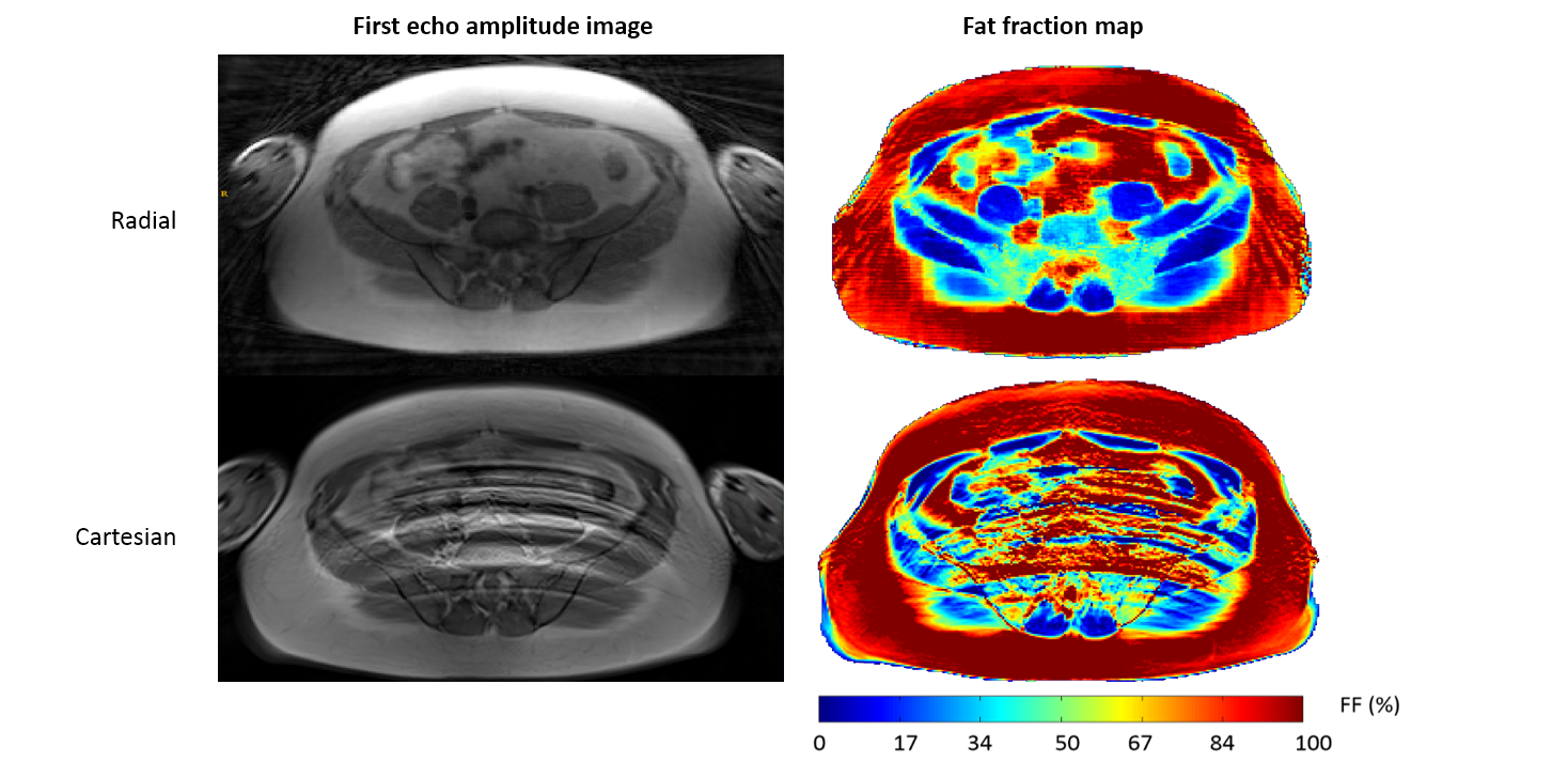
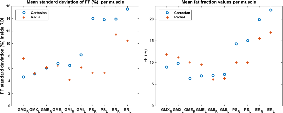
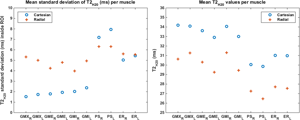
Figure 1 gives visual clues for the advantages of using a radial sequence: both the amplitude image and the fat fraction maps were strongly affected by the respiratory motion, with obvious artefacts particularly in the PS muscles. Those artefacts translate into an observable high standard deviation of FF and T2 measured inside these ROIs compared to muscles not affected by motion, such as the GMX. The standard deviations of parameters measured with the radial sequence did not depend on the muscle location, as can be seen on Figures 2 and 3. The precision of FF measurement was improved with the radial sequence, especially on the PS muscles which were heavily affected by motion artefacts, while a poorer precision of T2 estimation on the gluteal muscles was observed when compared to the Siemens sequence. The radial sequence provided comparable FF but significantly lower T2 values than the cartesian acquisition scheme. This difference might have its origin in more efficient gradient spoling in radial acquisition. Yet in the muscle affected by respiratory motion, FF was smaller with the radial scheme. The difference in standard deviation between motion affected muscles and non-affected muscles was significant for cartesian MSME.Discussion & Conclusion
These results showed that a radial multi-spin echo sequence allows water T2 and fat fraction estimations even in regions that are polluted by motion artefacts. In the scope of clinical applications, radial MSME implementation can be considered for upper body imaging, and for very young patients. Currently, the water T2 estimation is not as precise as with the Cartesian sequence but the precision is not impacted by motions artefacts. Future work will focus on improving the reconstruction algorithm and on using the parameter maps as a regularization criterion, eventually skipping raw image formation since the medical evaluation will be based on parametric images.Acknowledgements
This work was performed as part of the FP7 Bioimage-NMD projectReferences
1. Marty B, Baudin PY, Reyngoudt H, Azzabou N, Araujo ECA, Carlier P, de Sousa PL. Simultaneous Muscle Water T2 and Fat Fraction Mapping using Transverse Relaxometry with Stimulated Echo Compensation. NMR Biomed. 2016 Jan 27. doi: 10.1002/nbm.3459.
2. Fessler, J. A., & Sutton, B. P. (2003). Nonuniform fast Fourier transforms using min-max interpolation. IEEE Transactions on Signal Processing, 51(2), 560–574.
3. Zhang, T., Pauly, J. M., & Levesque, I. R. (2014). Accelerating parameter mapping with a locally low rank constraint. Magnetic Resonance in Medicine, 73(2), 655–661.
Figures