1425
Optimization of Reconstruction Parameters of Compressed Sensing STIR SEMAC for Metal Artifact Reduction MRI of Hip, Knee and Ankle Arthroplasty Implants: How many Iterations and how much Regularization is needed?1Johns Hopkins University, Baltimore, MD, United States, 2Siemens Healthcare GmbH, 3Siemens Healthcare USA
Synopsis
Compressed sensing-(CS)-based Slice Encoding for Metal Artifact Correction (SEMAC) turbo spin echo (TSE) pulse sequences achieve high–quality metal artifact reduction MRI around arthroplasty implants. Compressed sensing-based undersampling of k-space permits the time-neutral use of SEMAC, but requires iterative reconstruction algorithms, which are time consuming. We determined minimum number of iterations and regularization required for diagnostic image quality of STIR CS-SEMAC data sets of hip, knee and ankle arthroplasty implants. Our results show that 15-17 iterations and 0.0035 regularization results in optimal image quality of STIR CS-SEMAC images, which currently requires 4-5 minutes of reconstruction time.
Introduction
Compressed
sensing-(CS)-based acceleration for Slice Encoding for Metal Artifact Correction
(SEMAC) turbo spin echo (TSE) pulse sequence has been shown to be feasible for high–quality
metal artifact reduction MRI around arthroplasty implants [1-2]. When compared
to 3-fold accelerated conventional SEMAC TSE, an 8-fold-accelerated CS-SEMAC
pulse sequence can shorten the sampling time by up to 60% and reach acquisition
times that are similar to optimized, high-bandwidth TSE pulse sequences, thus
enabling the time neutral use of SEMAC [3]. CS-SEMAC reconstructions can be
performed inline; however, current reconstruction times for CS-SEMAC data sets
vary between 3-6 min depending on acquisition parameters such as number of
slices and spatial resolution, as well as reconstruction parameters such as the
number of iterations and regularization. A higher number of iterations improves
image clarity and higher regularization decreases image noise, but can be time
consuming. Thus, CS-SEMAC reconstructions should be performed with the minimum
number of iterations and regularization that is required to achieve diagnostic
image quality.Purpose
To optimize the length of time needed for image
reconstruction of STIR CS-SEMAC data sets of hip, knee and ankle arthroplasty
implants through the identification of the optimal minimum of the number of
iterations and regularization that is required for diagnostic image quality.Methods
This
study was approved by our institutional review board. Informed consent was obtained
from all participating subjects. Three subjects underwent 1.5T MR imaging
(MAGNETOM Aera, Siemens Healthcare, Erlangen, Germany) of the hip, knee and
ankle, using a 15-channel transmit-receive knee coil, 16-channel receive-only
ankle coil and 18-channel receive-only surface and embedded spine array coils
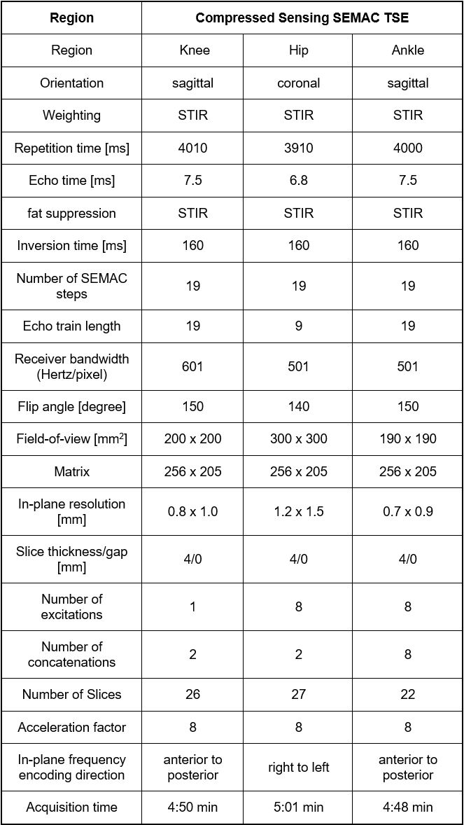
for hips. The study protocol consisted of 8-fold accelerated STIR CS-SEMAC pulse
sequences (Figure 1), which used 19 SEMAC-encoding steps and acceleration
through incoherent undersampling of the 2D-phase encoding matrix and
non-linear, SENSE-type reconstruction with L1-norm-based regularization was
used [4]. CS-SEMAC raw data of hip, knee and ankle arthroplasty implants were reconstructed
using 1, 3, 5, 7, 10, 13, 15, 17, 20, 30, 40 and 50 iterations and regularization
values of 0.0005. 0.0015, 0.0025, 0.0035, 0.0045 and 0.0055. To determine the
optimal minimum number of iterations that is required to achieve diagnostic image
quality, all possible combinations of iterations and regularization were created.
All reconstructed datasets were reviewed in consensus by two observers for
their diagnostic image quality taking into consideration the amount of implant-induced
susceptibility artifact as well as visibility of the implant-bone interface and
periprosthetic soft tissues. The reconstruction time for all image reconstructions
was recorded as well.Results
A total of 216 reconstructions of CS-SEAMC data of the hip,
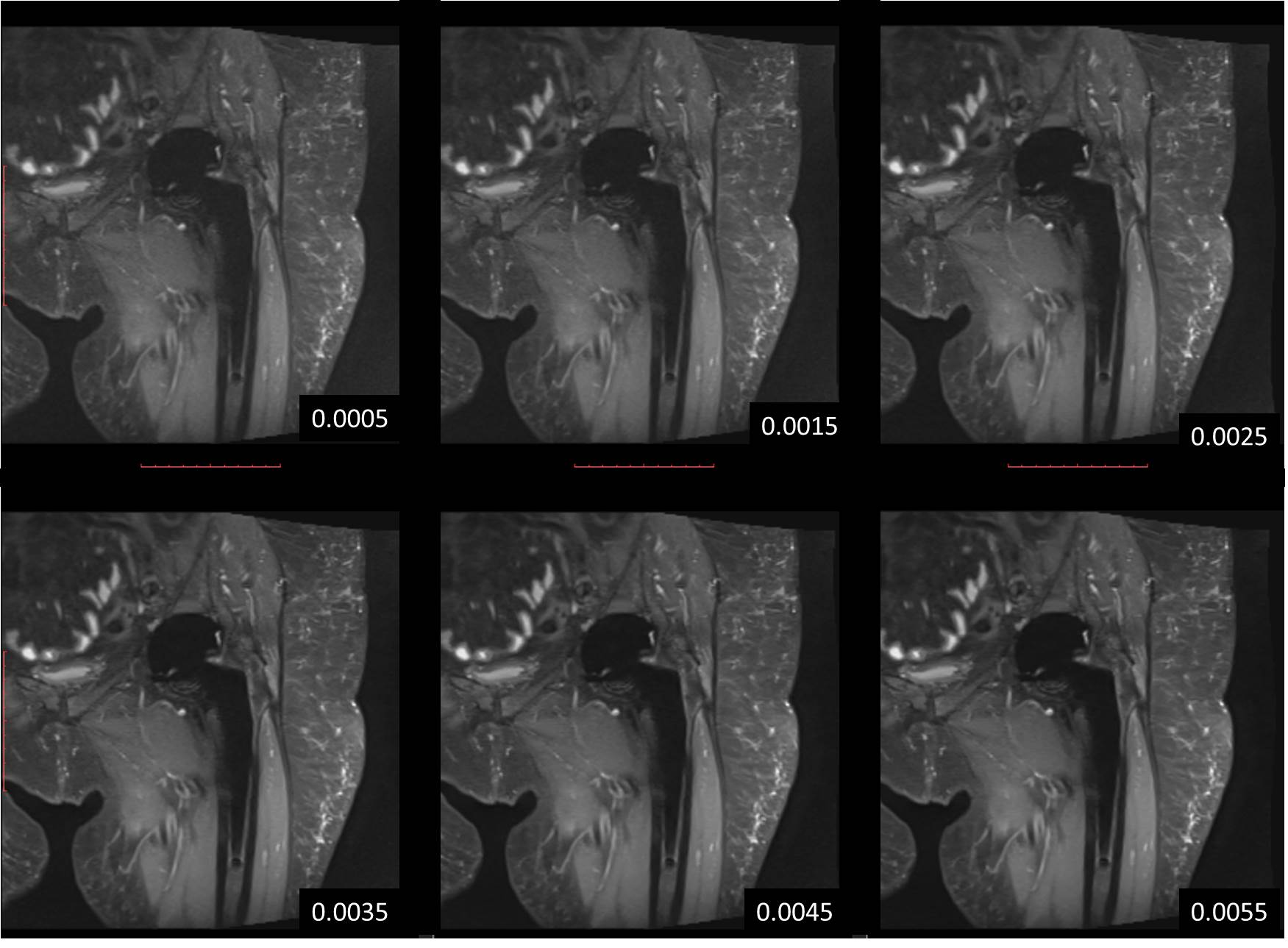
knee and ankle were successfully created and reviewed. Across all iterations, the
image quality achieved with a regularization of 0.0035 was ranked highest in
83.3% (10/12) of hip data sets, 100% (12/12) of knee data sets and 83.3%
(10/12) of ankle data sets, whereas higher regularizations did not result in
improving image quality, but resulted in edge blurring at regularization in
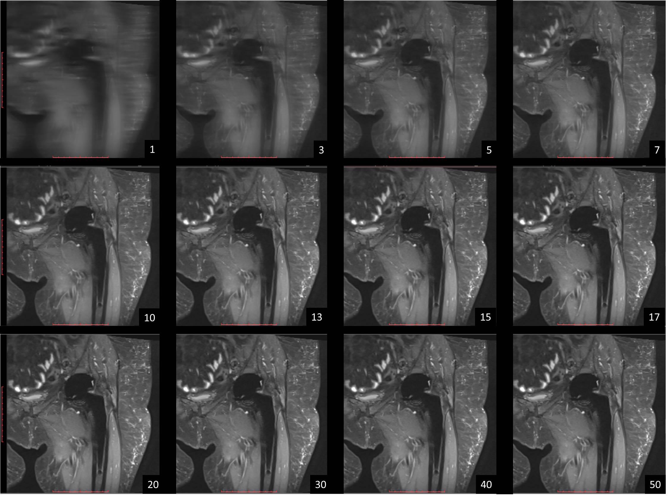
excess of 0.0035 (Figure 2). Across all regularization parameters, the image
quality achieved with 17 iterations was ranked highest in 66.7% (4/6) of hip
data sets and the image quality achieved with 15 iterations was ranked highest in
both 100% (6/6) of knee and 100% (6/6) of ankle data sets, whereas higher
numbers of iterations did not result in improving image quality (Figure 3). Image
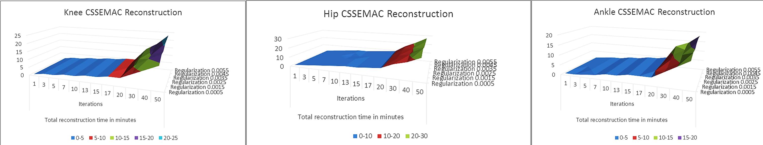
reconstruction times increased with increasing iterations ranging from <1 minutes
for 1 iteration to 25 minutes for 50 iterations (Figure 4).Discussion
The image quality of STIR CS-SEMAC data that were acquired through
pseudo-random undersampling, require iterative reconstruction that is dependent
on the number of iterations and degree of regularization. While the
reconstruction time is mainly a function of the number of iteration steps, our study
shows that beyond certain threshold numbers of iterations and regularization, no
additional gain in image quality may be achieved. The thresholds are 17
iterations for hip and 15 iterations for knee and ankle CS-SEMAC STIR data sets,
which require reconstruction times of 5 minutes, 4 minutes and 4 minutes
respectively. This has important practical implications as reconstruction
times are substantial for datasets of more than 20 iterations. Similarly, the
ideal regularization was found to be 0.0035 for STIR CS-SEMAC data sets.
Increasing the regularization beyond this threshold did not result in improving
image quality, but introduce blurring of contour with higher regularization.Conclusion
Optimal image quality of STIR CS-SEMAC MR images of hip,
knee and ankle may be achieved with regularization parameter of 0.0035 and minimum of 15-17 iterations, which has important practical implication to limit
the required reconstruction time. Acknowledgements
No acknowledgement found.References
1. Lu W, Pauly KB, Gold GE, Pauly JM, Hargreaves BA. SEMAC: Slice Encoding for Metal Artifact Correction in MRI. Magn Reson Med. 2009 Jul;62(1):66-76. doi: 10.1002/mrm.21967.
2. Fritz J, Ahlawat S, Demehri S, Thawait GK, Raithel E, Gilson WD, Nittka M. Compressed Sensing SEMAC: 8-fold Accelerated High Resolution Metal Artifact Reduction MRI of Cobalt-Chromium Knee Arthroplasty Implants. Invest Radiol. 2016 Oct;51(10):666-76
3. Fritz J, Fritz B, Thawait GK, Raithel E, Gilson WD, Nittka M, Mont MA. Advanced metal artifact reduction MRI of metal-on-metal hip resurfacing arthroplasty implants: compressed sensing acceleration enables the time-neutral use of SEMAC. Skeletal Radiol. 2016 Oct;45(10):1345-56.
4. Liu j, Rapin J, Chang T, Schmit P, Bi X, Lefebvre A, Zenge M, Mueller E, Nadar M. Regularized reconstruction using redundant Haar wavelets: A means to achieve high under-sampling factors in non-contrast-enhanced 4D MRA. ", Proceeding of the International Society for Magnetic Resonance in Medicine, 20TH Annual Meeting and Exhibition, Melbourne, Australia, 5-11 May 2012, Vol. 20, 21 April 2012 (2012-04-21), pages 2237.
Figures