1419
Accelerating 3D Head-and-Neck MR Imaging Using Compressed Sensing with Structure-Guided Total Variation for MR-Guided Multi-Fractional Radiotherapy1Medical Physics and Research Department, Hong Kong Sanatorium & Hospital, Happy Valley, Hong Kong, 2Department of Radiotherapy, Hong Kong Sanatorium & Hospital, Happy Valley, Hong Kong
Synopsis
MR image-guided radiotherapy (IGRT) holds potentials on outcome improvement in the head-and-neck (HN) radiotherapy. Patients receiving MR-guided multi-fractional HN IGRT are immobilized in each treatment fraction and set up to the exact position as of the treatment planning scan. Inter-fractional MR images are supposed to show highly correlated anatomy structure and edge information which can be incorporated into compressed sensing (CS) based MR reconstruction to shorten the scan time while preserve image quality in multiple fractions. In this study, we investigated the feasibility of accelerating high spatial resolution 3D MRI using CS with structure-guided total variation for multi-fractional HN radiotherapy.
Introduction
MR image-guided radiotherapy (IGRT) holds potentials on outcome improvement in the head-and-neck (HN) radiotherapy. Patients receiving MR-guided multi-fractional HN IGRT are immobilized in each treatment fraction and set up to the exact position as of the treatment planning scan to ensure accurate dose delivery. Therefore, inter-fractional MR images in the immobilized treatment position show highly correlated anatomical structure information. Such inherent structural correlation can be incorporated into the compressed sensing (CS) based MR reconstruction1 to shorten the scan time2-6 while preserve fine structural information. In this study, we aimed to investigate the feasibility of accelerating high spatial resolution 3D MR imaging using compressed sensing (CS) with structure-guided total variation (TV) for multi-fractional HN radiotherapy.The proposed method was tested using in-vivo human experiments under radiotherapy setup.Method
Image Acquisition under Radiotherapy Setup
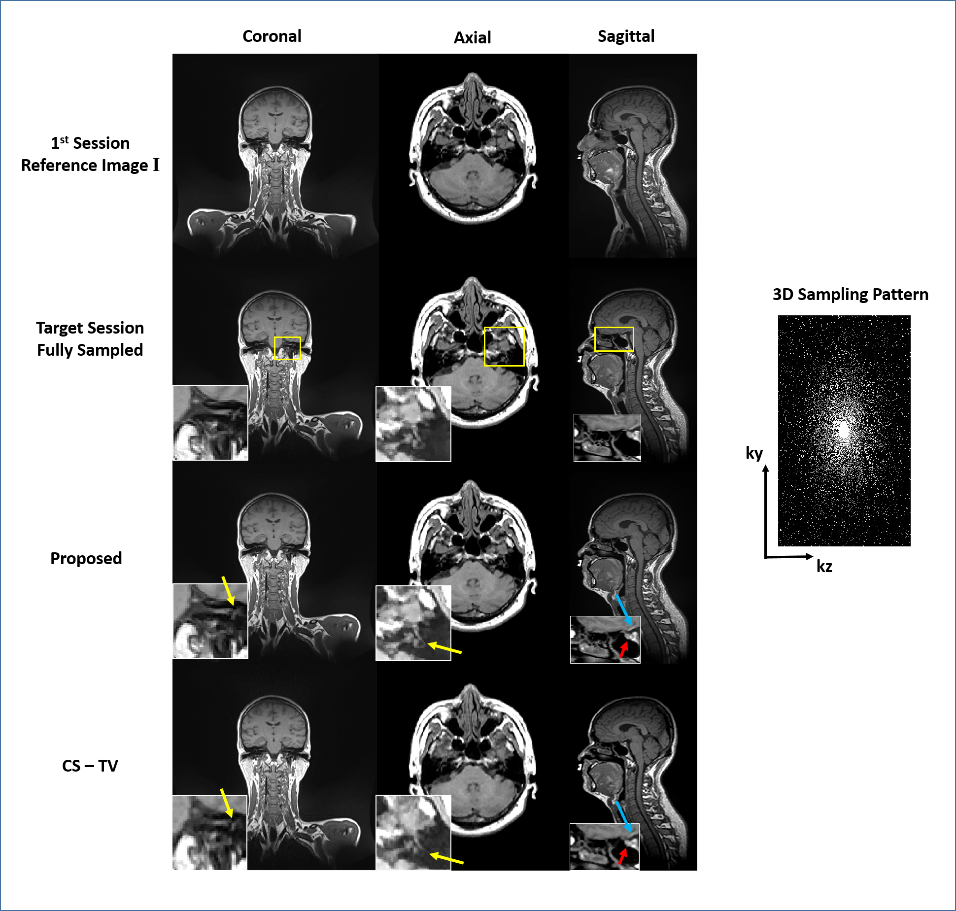
Eleven healthy volunteers were recruited and informed consent was obtained. MR images were acquired on a 1.5-Tesla MR scanner (Aera, Siemens Healthineers, Erlangen, Germany) dedicated for radiotherapy applications. Each volunteer received a series of scans (4-40 times) in an immobilized RT treatment position to simulate the HN-RT treatment fractions. All volunteers were immobilized using a personalized 5-point thermoplastic mask, and carefully aligned using a well-calibrated 3-dimensional external laser system. T1-weighted SPACE images were acquired with: FOV = 470mm×470mm×269mm and matrix size = 448×448×256 (PE×FE×SE); TR/TE = 420/7.2 ms, echo train length (ETL) = 40, bandwidth = 657Hz/pixel, yielding an isotropic voxel size of 1.05mm. The k-space was retrospectively undersampled along the phase-slice encoding direction using a 2D Poisson Disc sampling pattern with a reduction factor of 8. Images from the first scan were used as the anatomical prior.
Image Reconstruction
Following the conventional CS framework, the reconstruction of the i-th scan session images can be formulated as $$\mathop {\arg \min }\limits_{\bf{x}} \left\| {{\bf{Fx}} - {\bf{y}}} \right\|_2^2 + \lambda R({\bf{x}})\tag{1},$$ where $$${\bf{F}}$$$ is the undersampled Fourier transform, $$${\bf{y}}$$$ represents the acquired undersampled k-space data, $$$R({\bf{x}})$$$ is the regularization function, and $$$\lambda $$$ is the regularization parameter.
In multi-fractional radiotherapy, as the patient is scanned in the immobilized treatment position using the same imaging protocol, images acquired from multiple sessions are expected to show high correlation on anatomical structures and edges to the first scan session image. To exploit such inherent structural correlation, we proposed to use the three-dimensional structure-guided total variation as the penalty function to aid the reconstruction process. Specifically, instead of blindly and uniformly enforcing local smoothness on an image itself, the structure-guided total variation enforces the parallelity between the reconstructed image $$${\bf{x}}$$$ and the first scan reference image $$${\bf{I}}$$$’s gradients, or mathematically as $$R({\bf{x}}|{\bf{I}}) = \sum\limits_{n = 1}^N {{{\left( {{{\left| {\nabla {{\bf{x}}_n}} \right|}^2} - {{\left\langle {\nabla {{\bf{x}}_n},{{\bf{\xi }}_n}} \right\rangle }^2}} \right)}^{1/2}}} \tag{2},$$ where $$$N$$$represents the total number of pixels, $$$n$$$ is the pixel index, $$$\nabla $$$ denotes the three dimensional discrete gradient operation to compute the image gradient along three orthogonal directions, $$${{\bf{\xi }}_n} = \nabla {{\bf{I}}_n}/{\left| {\nabla {{\bf{I}}_n}} \right|_\tau }$$$ with $$${\left| {\nabla {{\bf{I}}_n}} \right|_\tau } = {({\left| {\nabla {{\bf{I}}_n}} \right|^2} + {\tau ^2})^{1/2}}$$$ denotes the normalized spatially varying gradient field obtained from $$${\bf{I}}$$$, and parameter $$$\tau $$$ penalizes the influence of noise. Such measure of structure similarity using the parallelism of paired images’ gradients is also known as a special case of asymmetric parallel level sets6 and has been proven to be convex that allows the employment of any smooth minimization methods.
Based on the abovementioned model, the compressed
sensing reconstruction with the structure-guided total variation
regularization can be formulated as $$\mathop {\arg \min }\limits_{\bf{x}} \left\| {{\bf{Fx}} - {\bf{y}}} \right\|_2^2 + \lambda R({\bf{x}}|{\bf{I}})\tag{3}.$$
The underlying convex optimization problem was
solved using alternating direction method of multipliers (ADMM).
Results
Figure 1 shows different views of one representative 3D image of a volunteer acquired from the first session (the reference image $$${\bf{I}}$$$), the fully sampled images of targeted session and the reconstructed image using the proposed method. Conventional CS with TV (CS-TV) was performed for comparison. In general, the proposed method accurately reconstructed the images from highly undersampled k-space while CS-TV showed noticeable blurring and aliasing artifacts. In particular, the proposed method better preserved the fine structures of important small organs-at-risks (OARs) in HN radiotherapy such as optical chiasm, pituitary gland and inner ear than CS-TV.Conclusion
This pilot study investigated the feasibility to accelerate the image acquisition for MR-guided multi-fractional head-and-neck radiotherapy using compressed sensing with structure-guided total variation regularization. The preliminary results showed that the proposed method could preserve spatial details of fine OAR structures at highly accelerated scans. Further thorough validation on real HN cancer patient data is warranted.Acknowledgements
No acknowledgement found.References
1. Lustig M, Donoho D, Pauly JM. Sparse MRI: the application of compressed sensing for rapid MR imaging. Magn Reson Med 2007;58:1182–1195.
2. Li Y, Dumoulin C. Correlation imaging for multiscan MRI with parallel data acquisition. Magn Reson Med 2012;68:2005–2017.
3. Gong, E, Huang, F, Ying, K, Wu, W, Wang, S and Yuan, C, PROMISE: Parallel-imaging and compressed-sensing reconstruction of multicontrast imaging using SharablE information. Magn. Reson. Med. 2015, 73: 523–535.
4. Bilgic B, Goyal VK, Adalsteinsson E. Multi-contrast reconstruction with Bayesian compressed sensing. Magn Reson Med 2011; 66:1601–1615.
5. Peng X, Ying L, Liu Q, Zhu Y, Liu Y, Qu X, Liu X, Zheng H and Liang D, Incorporating reference in parallel imaging and compressed sensing. Magn. Reson. Med. 2015, 73: 1490–1504. doi:10.1002/mrm.25272
6. Ehrhardt MJ, Markiewicz P, Liljeroth M, Barnes A, Kolehmainen V, Duncan JS, Pizarro L, Atkinson D, Hutton BF, Ourselin S, Thielemans K, Arridge SR. PET reconstruction with an anatomical MRI prior using parallel level sets. IEEE Trans Med Imaging. 2016, 35(9):2189-2199.
Figures