1404
Contrast and Resolution Mixing for Magnetic Resonance Fingerprinting1Application Development, Siemens Healthcare, Erlangen, Germany, 2Friedrich-Alexander Universität Erlangen-Nürnberg, Erlangen, Germany, 3Department of Biomedical Engineering, Case Western Reserve University, OH, United States, 4Dept. of Radiology, Case Western Reserve University and University Hospitals of Cleveland, OH, United States
Synopsis
Quantitative parameter maps obtained from Magnetic Resonance Fingerprinting (MRF) are sensitive to B1+ inhomogeneities. Extending a dictionary by an additional B1+ dimension is a promising approach to account for this. In order to improve the differentiation of data in the B1+ dimension, we implemented a novel B1+ sensitive encoding. This approach employs the extension of the conventional FISP encoding by RF spoiled parts. Since high undersampling factors prevent a direct implementation of this technique, the undersampling pattern is varied during the acquisition. We use a spiral acquisition scheme which samples most of the FISP encoded parts of the fingerprint with high resolution. RF spoiled parts as well as a small fraction of the FISP encoded parts are being sampled with low resolution.
Purpose
Magnetic Resonance Fingerprinting (MRF) 1 is a new technique that promises multi-parametric, quantitative MRI. Spatial variation of the RF transmit field (B1+) is a commonly observed effect, that may lead to systematic errors in MRF results 2,3. A straightforward approach would incorporate the magnitude of the B1+ field as a separate dimension into the dictionary. However, also a dictionary with a B1+ dimension is vulnerable for mismatches in case of the additional dimension.
For
the purpose of a more reliable assignment of quantitative parameters, we
propose a novel encoding scheme for MRF, that adds additional B1+ dependent segments
to the signal evolution. This is accomplished by a combination of changing
the encoding scheme and the usage of sampling patterns with different
resolutions.
Methods
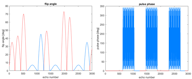
We use a prototype implementation of the MRF method from 4, which employs a fast imaging with a steady-state precession (FISP) sequence with temporal variation of repetition time and flip angle. This established encoding pattern is extended by RF-spoiled parts with low flip angle. Figure 1 shows the novel encoding.
These parts are characterized by low flip angles, quadratic phase increment from one RF pulse to the next and take longer than the FISP segments (compare figure 1 for visualization of used flip angles and pulse phase). The MR signal generated by these FLASH segments varies slowly and is mainly B1+ and T1 dependent. Since RF spoiling destroys higher-order coherences, the T2 sensitivity of the signal is negligible. The FISP segments following the RF-spoiled parts benefit from high remaining magnetization and thus enable better differentiation in the T1 and T2 dimension with higher SNR.
Theoretical and experimental work has shown that especially the FLASH encoding is very sensitive to undersampling artifacts. To account for this, a low-resolution spiral acquisition, for example with an undersampling factor of 11, is required. Another promising strategy is to combine highly undersampled FISP segments and little undersampled RF-spoiled parts. For robust B1+ mapping, it is necessary that also a small part of the FISP segments is little undersampled.
For the evaluation of this method, human brain scans were performed on a clinical 3T MR scanner (MAGNETOM Skyra, Siemens Healthcare, Erlangen, Germany).
Results
Figure 2 shows results obtained with the new B1+ sensitive encoding on a human brain (field of view of 300mm, undersampling factor 11, resolution of 2.3 m, slice thickness 5mm). The whole fingerprint was acquired with this setting, i.e. the resolution remained the same during the acquisition. The dictionary’s relative B1+ values range from 75% to 125% in 5% steps.
Keeping field of view and slice thickness, but increasing the undersampling to a factor of 48 and the resolution to 1.2 mm, leads to parameter maps as shown in figure 3a).
Parameter maps from combined fingerprints, consisting of signal parts from a high-resolution acquisition (FoV 300mm, resolution 1.2mm, slice thickness 5mm, undersampling factor 48) and signal parts from a low-resolution acquisition (FoV 300mm, resolution 2.3mm, slice thickness 5mm, undersampling factor 11) as described in figure 1 are shown in figure 3b).
Discussion
The parameter maps obtained with the novel encoding in the human brain with a resolution of 2.3*2.3*5.0 mm3 are smooth for B1 as well as for T1 and T2. Parameter maps with higher resolution are artifact afflicted, especially the relative B1 and T2 maps. The ones from combined signals are free of artifacts by visual inspection and come without time and resolution penalty as compared to traditional FISP-based MRF.
It is possible to add a B1+ dimension to the dictionary by using FLASH segments in the encoding. This introduces limitations regarding resolution and undersampling factor in order to ensure stable pattern matching. These limitations can be overcome by varying the undersampling throughout the acquisition of the fingerprints. Our implementation, combining highly undersampled FISP and little undersampled FLASH segments, shows the practicability of this technique. It produces artifact-free parameter maps with clinically feasible resolution without any scan time prolongation.
Conclusion
We demonstrate a novel method to better discriminate entries of an MRF dictionary including the B1+ field as an additional dimension. Results show that this can be achieved by using a combination of different signal evolution segments with adapted resolutions. These different signals do not need to be acquired in separated segments and provide time-efficient sampling by reducing dead times. Unlike other B1+ encoding strategies for MRF (e.g. 5), all signal is used for matching all parameters. The presented implementation does not take longer than a conventional FISP MRF scan and exhibits similar T1 and T2 differentiation quality.
Acknowledgements
No acknowledgement found.References
Ma D. et al., Magnetic Resonance Fingerprinting, Nature 2013
Jiang Y. et al. Simultaneous T1 and T2 Quantitation of the Human Brain at 7 Tesla by MR Fingerprinting ISMRM 2015
Chen Y. et al, Magnetic Resonance Fingerprinting (MRF) for Rapid Quantitative Abdominal Imaging, Proceedings of the ISMRM 2014.
Jiang Y. et al, MR fingerprinting using fast imaging with steady state precession (FISP) with spiral readout, MRM 2014
Buoncontrini et al., MR fingerprinting with simultaneous B1 estimation, MRM 2016
Figures
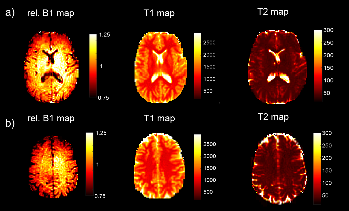
Parameter maps from a spiral B1 sensitive MRF acquisition of a human brain. Spiral was designed to cover an isotropic field of view of 300 mm with a resolution of 2.3 mm. a) and b) show two slices of the same brain.
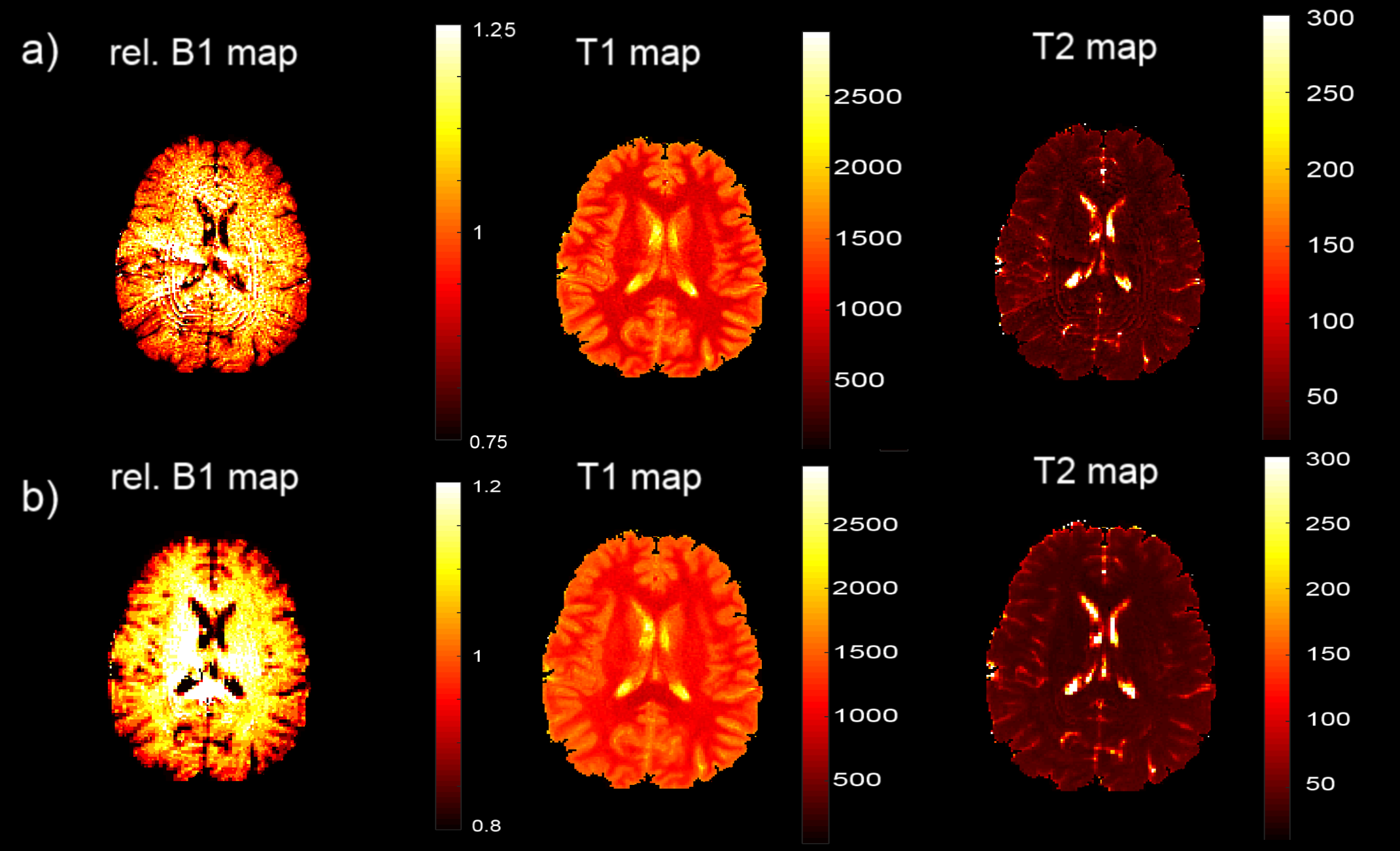
Parameter maps from a spiral B1 sensitive MRF acquisition of a human brain. a) One spiral with field of view of 300 mm and a resolution of 1.2 mm was used to sample the signal. b) Two different spiral designs with a field of view were used. For the high resolution parts a spiral with a resolution of 1.2 mm and for the low Resolution parts a spiral with a resolution of 2.3 mm was used.