1398
Shimming for BOLD sensitivity in the brain1Oxford Centre for Functional MRI of the Brain, University of Oxford, Oxford, United Kingdom
Synopsis
Whole brain resting state fMRI benefits greatly from shimming due to increases in BOLD sensitivity. This work presents a rapid shim calculation method using prior knowledge of nonlinear optimization results on a large database of field maps, which can significantly reduce the time spent on shim determination for BOLD sensitivity optimization, whilst delivering shim quality that is closer to the results of nonlinear optimization.
Introduction
Typically, a linear least-squares approach is used to calculate the shim currents that are required to minimize the standard deviation of the field inhomogeneity. Other methods also exist including, for example, minimization of the gradient of the local field or maximization of blood-oxygen-level dependent (BOLD) sensitivity.1,2 However, these complex, nonlinear optimization algorithms are not often used due to time constraints and local minimum challenges. Here we propose a rapid shim calculation method using prior knowledge of nonlinear optimization results generated based on a large database of field maps. This information can then be used in the linear optimization to deliver shim quality that is closer to the results of nonlinear optimization. The performance of our method in improving BOLD sensitivity was evaluated in comparison to the standard deviation (STD) optimization with linear least squares method and the direct BOLD sensitivity (BOLD-S) optimization with nonlinear least squares.Methods
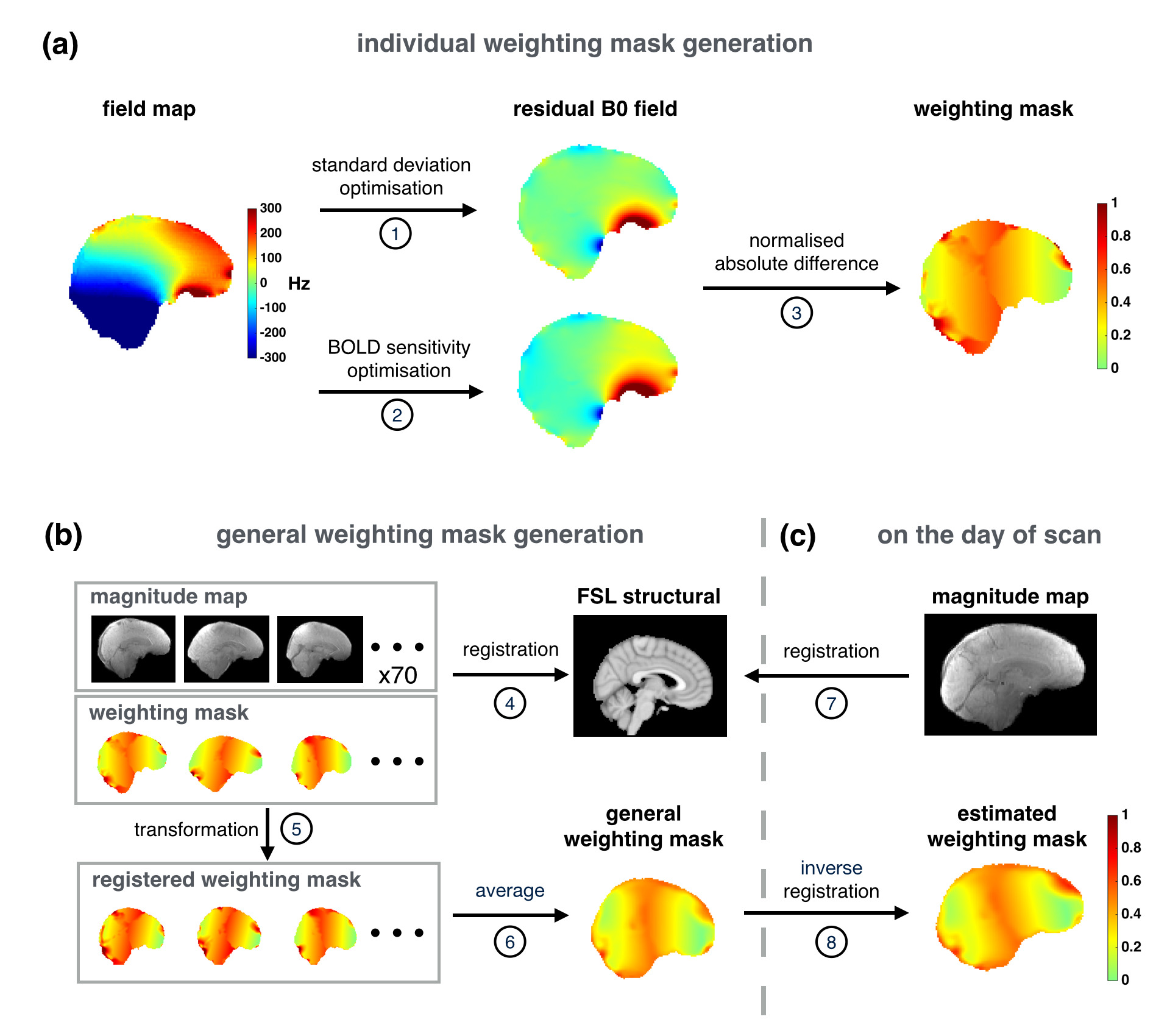
Experiments were performed on a Siemens 7T MRI scanner (Erlangen, Germany). Whole-brain B0 mapping was performed on 70 subjects in vivo with an asymmetrical dual gradient-echo based field mapping sequence (voxel size=2mm*2mm*2mm, TE1 =4.08ms, ΔTE=1.02ms). The proposed method uses a weighting mask in the standard deviation optimization (STD-WM). The choice of weighting is determined by identifying regions where improvements in field homogeneity translate into improvements in the BOLD sensitivity. The method is summarized in Figure 1.
Generation of weighting masks for all the subjects in the database
1. Optimize the field homogeneity by minimizing the standard deviation.
2. Optimize the field homogeneity for maximizing simulated BOLD sensitivity using a nonlinear least squares algorithm based on routine EPI acquisition settings (voxel size=2mm*2mm*2mm, TE=25ms, bandwidth=1578Hz, echo spacing=72ms). In order to guarantee satisfactory signal intensity and geometric distortion over the brain, residual B0 field standard deviation was limited to not greater than 120% of the STD method and the mean gradient in the phase-encoding direction was not greater than 1.5Hz/pixel (5% compression of geometric distortion).3
3. Take the absolute value of field maps optimized with the STD and BOLD-S methods, subtract absolute deviation maps optimized by BOLD-S to STD and normalize their differences to generate a weighting mask for a subject (lower values give higher weights).
Generation of a general weighting mask
4. Register the field map acquisition to standard space using FLIRT.4
5. Transform weighting masks to standard space using the registration transformation matrices generated in step 4.
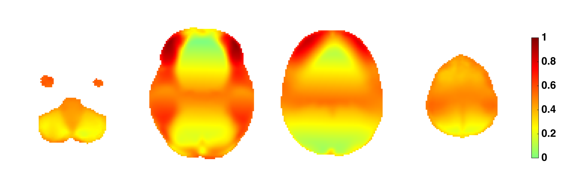
6. Average weighting masks in standard space to create a general weighting mask (Figure 2).
Fast optimization of shim settings on the day of scan
7. Acquire a field map of the scanned subject and register to standard space.
8. Apply the inverse registration to the general weighting mask to estimate a weight mask for the scanned subject.
9. Calculate the shim currents which minimize the standard deviation of the field map along with the weighting mask.
To evaluate the method, a leave-one-out cross validation technique was implemented to assess the shimming performance of three methods: the proposed method, the standard deviation optimization and the BOLD sensitivity optimization.
Results
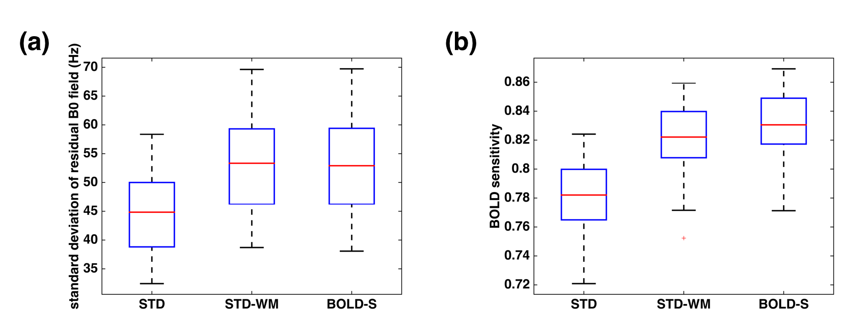
Figure 3 compares the simulated shimming results using the STD, the STD-WM and the BOLD-S optimization methods. Across all the subjects in database, the BOLD-S method improved the whole brain BOLD sensitivity in comparison to the STD method. The STD-WM method produced BOLD sensitivity improvements close to the BOLD-S method, but without significantly compromising B0 standard deviation.
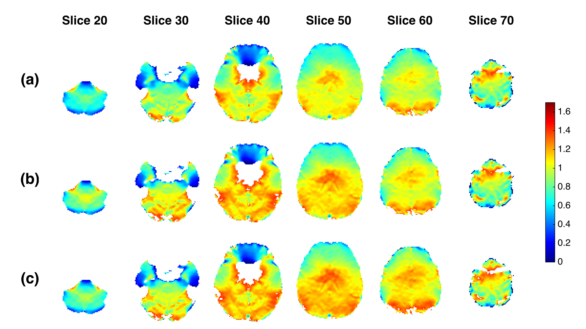
In Figure 4, it is evident that the STD-WM method improved BOLD sensitivity for example slices, comparing to the STD method. Meanwhile, the STD-WM method delivered similar pattern of results as the BOLD-S method, but without time penalty on shim calculation.
Discussion
The choice of target for shimming optimization depends on the application, but the increase in popularity of whole brain resting state fMRI motivated selection of a method that optimizes BOLD sensitivity. Our proposed STD-WM method identifies those regions where targeting field uniformity will increase overall BOLD sensitivity across the whole brain and accelerate the shim determination for BOLD sensitivity optimisation. The results using the STD-WM method produce BOLD sensitivity improvements that were similar to those from the BOLD optimization method and outperform those results from the STD method. However, one challenge of the STD-WM method is its dependence on specific EPI parameters. Any changes in EPI parameters will require the optimization to be rerun over the whole database.Conclusion
The STD-WM method significantly reduces the shim calculation time for BOLD sensitivity optimization and provides comparable results to direct BOLD sensitivity optimization.Acknowledgements
No acknowledgement found.References
1. Deichmann R, Josephs O, Hutton C, Corfield DR, Turner R. Compensation of susceptibility-induced BOLD sensitivity losses in echo-planar fMRI imaging. Neuroimage. 2002;15:120–135
2. Balteau E, Hutton C, Weiskopf N. Improved shimming for fMRI specifically optimizing the local BOLD sensitivity. Neuroimage. 2010;49(1):327-36.
3. Jezzard P, Clare S. Sources of distortion in functional MRI data. Hum Brain Mapp. 1999;8:80–85
4. Jenkinson M, Bannister P, Brady JM and Smith SM. Improved Optimisation for the Robust and Accurate Linear Registration and Motion Correction of Brain Images. NeuroImage. 2002;17(2):825-841.
Figures