1383
Validation of MRE measurements of shear modulus in both annulus fibrosus and nucleus pulposus within in two animal species1Mechanical Engineering, Polytechnique Montreal, Montreal, QC, Canada, 2Research Center, Sainte-Justine Hospital, Montreal, QC, Canada, 3NeuRA, Sydney, Australia
Synopsis
Magnetic resonance elastography (MRE) is an effective method to measure the shear modulus of the nucleus pulposus, and its changes with degenerescence. However, MRE was not used for the annulus fibrosus. This study validated the use of MRE for the measurements of the shear modulus variations within the intervertebral discs between regions (annulus fibrosus versus nucleus pulposus) and animal species (bovine versus kangaroo). Shear dynamic mechanical tests and MRE showed equivalent tissue differences. This study also highlighted that the loading history of the intervertebral disc has to be considered when choosing an animal model.
Purpose
The intervertebral disc main role
is mechanical, transmitting and distributing forces produced by the body weight
and spinal muscles activity. The mechanical properties of both the annulus
fibrosus and nucleus pulposus are altered by degenerescence (1).
Magnetic resonance elastography (MRE) is an effective method to measure the shear
modulus of the nucleus pulposus (2,3,4), and is sensitive to tissue
degenerescence (2). However, MRE was not applied to the annulus
fibrosus because of its small thickness and high stiffness that requires challenging
measurements at high frequencies. The objective of this study was to validate
MRE for the measurements of shear modulus variations within the intervertebral
discs between regions and animal species.Methods
Intervertebral joints of kangaroo and bovine tails were isolated. Bovine caudal discs are often used to study intervertebral disc degeneration because of similar size, biochemical composition and structure as human discs. However, the kangaroo caudal discs are weight bearing as the animal leans on his tail to walk (5). Because of different loading histories, bovine and kangaroo discs may present different shear moduli. Surrounding muscles and ligaments tissues were removed, and the intervertebral disc attached to the proximal vertebra was kept for MRE. MRI measurements were performed on a 9.4T Bruker system with a 50mm volume coil. The protocol was composed of high resolution anatomy sequence (Turbo-RARE-T2, TE=11.6ms, TR=2485ms, resolution 0.01mmx0.01mm) and a MRE spin echo sequence (mSME-MRE, TE=21.25ms, TR=1875ms, resolution 0.3mmx0.3mm, 9 slices, 0.3mm thickness, no gap) with sinusoidal motion encoding gradients synchronized to the vibration (frequency of 1200Hz). Shear dynamic mechanical tests were carried out on samples of annulus fibrosus and nucleus pulposus of bovine and kangaroo discs using a rheometer (Kinexus, Pro, Malvern instruments) equipped with serrated flat plattens. Samples were compressed with a 0.5N axial force in order to ensure full contact and allowed to equilibrate. A dynamic frequency sweep was performed with logarithmically spaced frequencies from 0.1Hz to 10Hz. The dynamic shear moduli G* were computed using custom MRE software (6) and R-space software (Malvern instruments, version 1.61) and compared between species and regions using a two-ways analysis of variances.Results
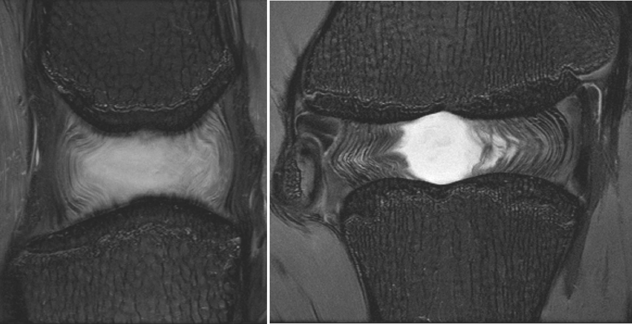
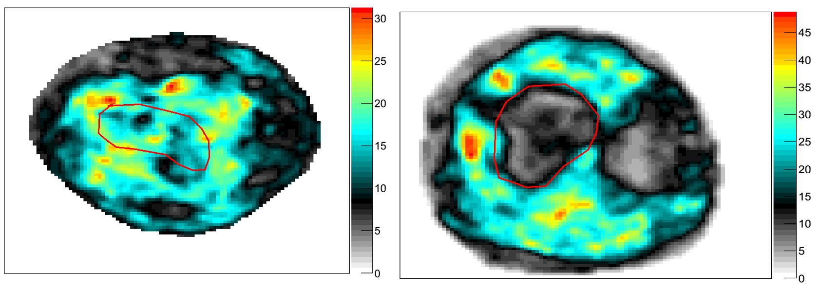
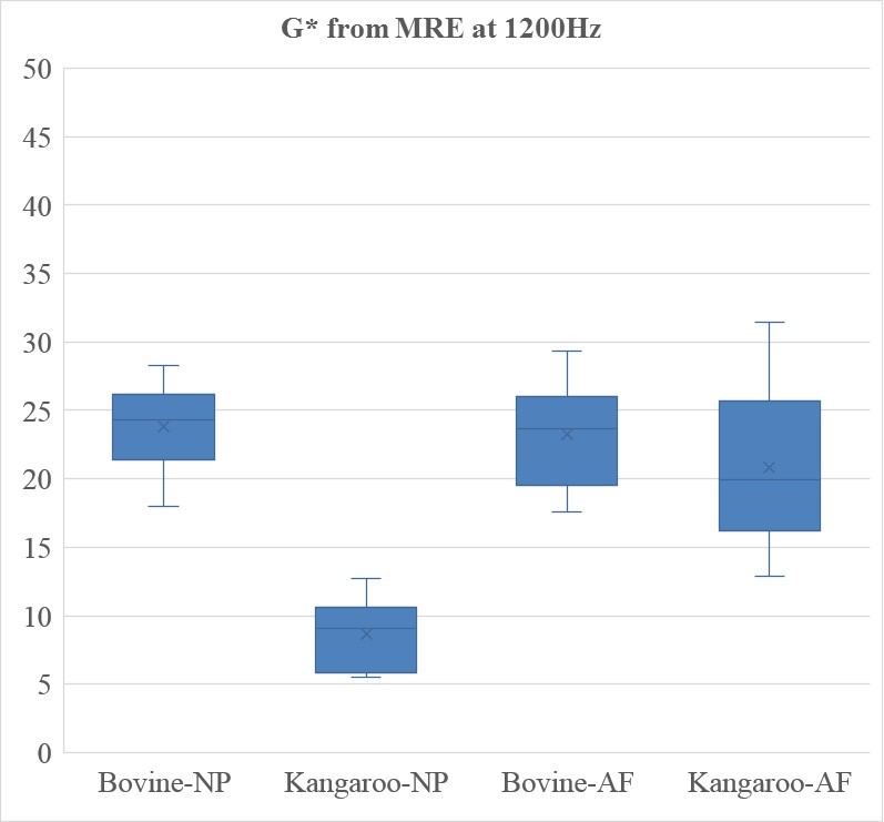
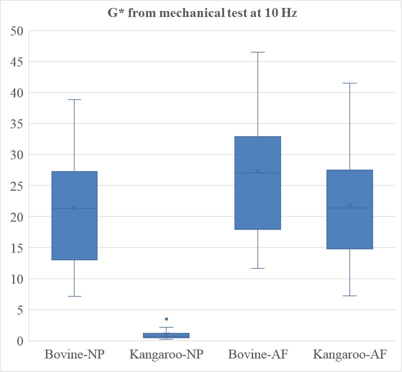
The kangaroo nucleus pulposus was more gelatinous than the bovine while the kangaroo collagen fibers of the inner annulus fibrosus seemed more organised (Figure 1). G* from MRE (Figure 2) decreased in the kangaroo disc from 20.9±6.5kPa in the annulus fibrosus to 8.7±2.7kPa in the nucleus pulposus, and remained constant in the bovine disc at 23.2±4.1kPa in the annulus fibrosus and 23.8±3.4kPa in the nucleus pulposus (Figure 3). G* from the dynamic shear test at 10Hz decreased in the kangaroo disc from 21.9±10.3kPa in the annulus fibrosus to 1.0±0.4kPa in the nucleus pulposus, and decreased slightly in the bovine disc from 27.3±10.1kPa in the annulus fibrosus to 21.4±7.0kPa in the nucleus pulposus (Figure 4). For both mechanical and MRE results, significant differences were observed between species and between regions (p<0.001), but not between the bovine annulus fibrosus, the bovine nucleus pulposus and the kangaroo annulus fibrosus (p>0.05).Discussion
The dynamic shear modulus we measured within the bovine nucleus pulposus was smaller but in the same range than the one measured at 1000Hz in the baboon (79±15 kPa)3, 10 to 30 times smaller than the one measured at 1250Hz in the human (661 kPa, interquartile range 234-980)2 and higher but in the same range than the one measured at 50Hz in the bovine (6.51 ± 1.27 kPa)4. The softer kangaroo nucleus pulposus was in agreement with its more gelatinous consistency than the bovine nucleus pulposus. The kangaroo nucleus pulposus might have higher proteoglycan content than bovine or human and thus might sustain higher hydrostatic pressure. The MRE procedure we chose, with a circular platen vibrating against the axial disc surface, simulated the vibration of the adjacent bone for further in vivo applications, and allowed a good wave propagation through the tissue. We did not compare the dynamic shear modulus from the two methods as the applied frequency was too different (10Hz versus 1200Hz).Conclusion
Both mechanical tests and MRE highlighted differences within the kangaroo discs between annulus fibrosus and nucleus pulposus, which validate the use of high frequency MRE within the annulus fibrosus and confirm the sensitivity of MRE to tissue mechanical differences. This study also highlighted that the loading history of the intervertebral disc has to be considered when choosing an animal model.Acknowledgements
The authors would like to thank André Bongers
for his help at the MRI facilities. This work was supported by the "Fond
de Recherche du Quebec en Nature et Technologies" (FRQNT, establishment of
new investigators) and the "Natural Sciences and Engineering Research
Council" (NSERC, discovery grant) of Canada.
References
1. Périé D., MacLean J.J., Owen J., Iatridis J.C. Effect of proteoglycan and water contents on the compressive modulus and permeability of the nucleus pulposus and annulus fibrosus. Annals of Biomedical Engineering 2006, 34(5):769-77. 2. Cortes D, Magland J, Wright A, Elliott D. The Shear Modulus of the Nucleus Pulposus Measured Using MR Elastography: A Potential Biomarker for Intervertebral Disc Degeneration. Magn Reson Med. 2014, 72(1): 211–219. 3. Ben-Abraham EI, Chen J, Felmlee JP, Rossman P, Manduca A, An KN, Ehman RL. Feasibility of MR elastography of the intervertebral disc. Magn Reson Imaging. 2015 doi: 10.1016/j.mri.2015.12.037. 4. Streitberger KJ, Diederichs G, Guo J, Fehlner A, Hamm B, Braun J, Sack I. In vivo multifrequency magnetic resonance elastography of the human intervertebral disk. Magn Reson Med. 2015 Nov;74(5):1380-7. 5. Alini et al. (2008). Are animal models useful for studying human disc disorders/degeneration? Eur Spine J 17:2–19. 6. Sinkus et al. (2000). High-resolution tensor MR elastography for breast tumour detection. Physics in Medicine & Biology 45: 1649-1664.Figures