1381
Integration of MR Elastography and Fat/Water Separation Imaging1Radiological Science, Tokyo Metropolitan University, Tokyo, Japan, 2National Institute of Advanced Industrial Science and Technology, Tsukuba, Japan, 3Mechanical Engineering, TOKYO DENKI UNIVERSITY, Tokyo, Japan, 4Judo Neurophysiotherapy, University of Toyama, Toyama, Japan, 5System Emotional Science, University of Toyama, Toyama, Japan
Synopsis
In the present study, we developed a method to combine a simple MRE technique with a fat/water separation method (Dixon method) based on a conventional gradient-echo type multi-echo MR sequence (GRE-MultiEcho-MRE). Because the proposed method used the GRE-multiecho MRE, it is possible to select shortest in-/opposed-phase TE in the 1st TE. Thus, the proposed method allows to increase signal-to-noise ratio of fat/water images.
PURPOSE
Fat is often a source of problems in MR imaging. It tends to have high signal intensity at all contrasts, which could mask image changes reflecting various tissue pathologies. It also causes two types of artifacts due to its structure, known as a chemical shift artefact and a phase cancellation artifact. Since these artifacts similarly affect the MR elastography (MRE), a fat suppression technique is required in all MRE sequences. In the present study, we developed a method to combine a simple MRE technique with a fat/water separation method (Dixon method) based on a conventional gradient-echo type multi-echo MR sequence (GRE-MultiEcho-MRE).BACKGROUND
Our previous study reported a new method for MRE using a conventional gradient-echo type multi-echo MR sequence without motion encoding gradient (MEG)1. In the all MRE, MR images must be taken in synchronization with some vibration phases. In the GER-MultiEcho-MRE system, the vibration phase offset was controlled by waveform generator, giving continuous (steady state) vibrations throughout the whole acquisition (each imaging period). The GER-MultiEcho-MRE uses a series of echoes acquired as a train following after a single excitation pulse. The multiple symmetrical gradient-echoes in the GER-MultiEcho-MRE are acquired by symmetrical bipolar readout gradient lobes. This readout gradient lobes (GRs) have a function similar to MEG (MEG-like effect). If GRs are synchronized with the vibration frequency, the later generated echo induces a greater MEG-like effect.
Dixon suggested in 1984 that in-phase echo time (TE) and opposed-phase TE images could be combined so that fat only and water only images could be created. The idea is that in the in-phase TE images, the signal (Sin) is the sum of fat (Sf) and water (Sw) signals (Sin=Sw+Sf), while, in the opposed-phase TE images, it (Sopp) is the difference (Sopp=Sw-Sf). By adding the two images, only the water signals remain, while the subtraction of the opposed-phase TE image produces a fat-only image.
Trzasko et al. (2015) reported a new sequence that combines a dedicated MRE (built-in MEG) and Dixon method2. In this study, we combined a Dixon method with the GRE-MultiEcho-MRE.
METHODS
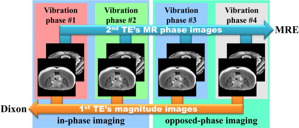
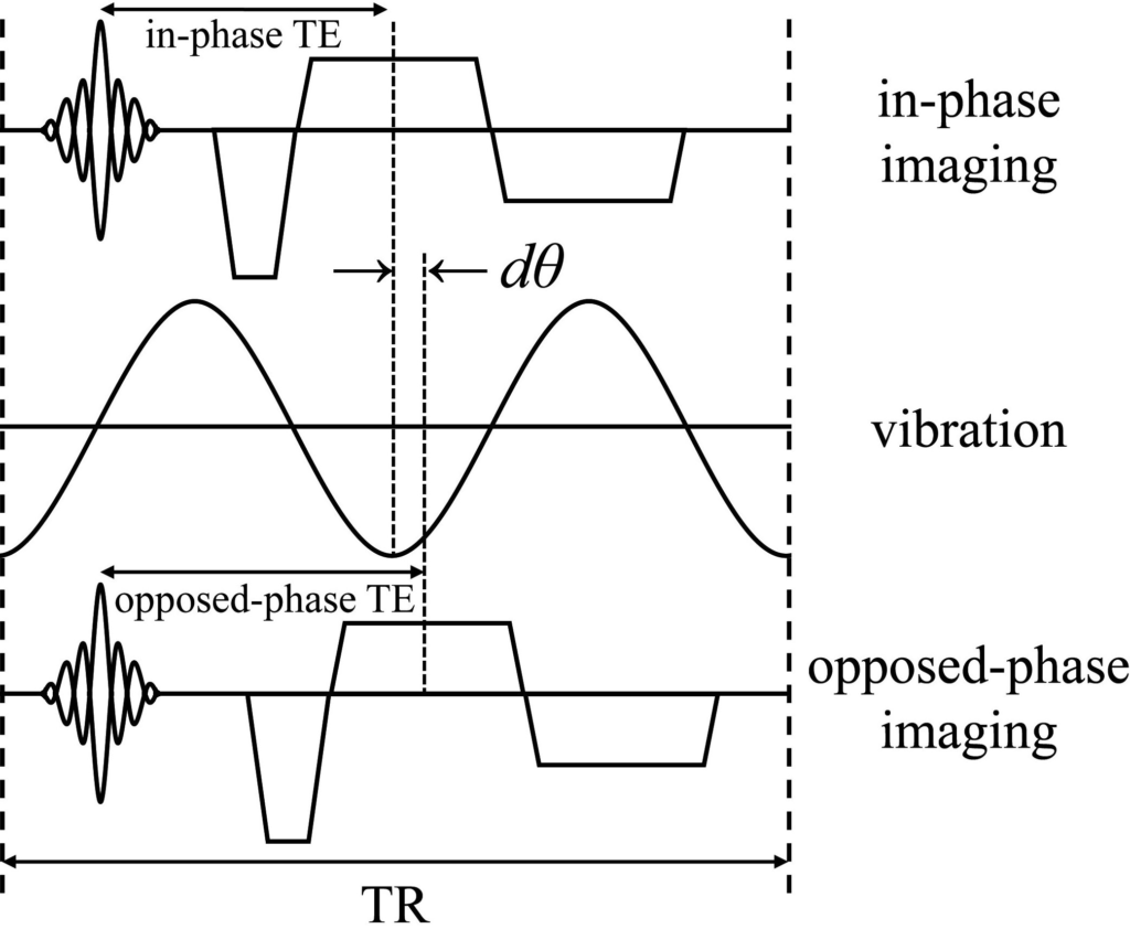
Figure 1 shows the image processing flow of the proposed method. In the GER-MultiEcho-MRE, the magnitude image of the 1st echo has the smallest MEG-like effect, but provides the best anatomical structure image because the 1st echo has the highest signal to noise ratio (SNR), and it is less affected by magnetic susceptibility artifacts. Therefore, in the proposed method, we applied the 1st echo (TE) images to Dixon method. The MR phase images of later echo (2nd TE) were used for MRE. The timing of the RGs was changed along with the changes in the TE; the vibration phase also changed (Fig.2). The TE of in-phase imaging and the TE of opposed-phase imaging were different, which causes vibration phase mismatch (dθ). This dθ was corrected by vibration waveform generator.
All MRE experiments were performed on a clinical MR imager (Achieva 3.0 T; Philips Healthcare, Best, The Netherlands) using a torso coil. A self-made waveform generation system (LabVIEW, USB-6221; National Instruments, TX, USA) was used to generate the vibration waveform. This system is capable of generating sinusoidal waveforms with arbitrary frequencies and phases. Power amplifier (XTi 1000; Crown, IN, USA) and a pneumatic pressure generator (Subwoofer TIT320C-4 12”; Dayton Audio, OH, USA) units were used to supply vibrations to a vibration pad. The vibration pad was designed using a three-dimensional (3D) printer (3D touch; 3D system, SC, USA) in order to adapt to buttock shapes.
The MRE sequence parameter were TR: 40ms, 1st TE: 2.2ms (in-phase) / 3.3ms (opposed-phase), 2nd TE: 12.2ms (in-phase) / 13.3ms (opposed-phase), flip angle: 20degree, matrix: 256×256, vibration frequency: 50Hz, vibration phase offset: 4, total acquisition time: 82s, MEG-like effect direction: L-R. All elastograms were produced by Local Frequency Estimate (LFE) algorithm freeware (MRE/Wave, MAYO CLINIC).
RESULTS and DISCUSSION
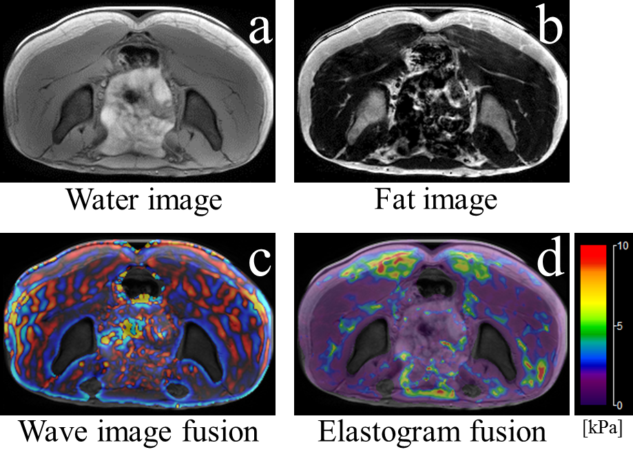
Figure 3 shows the water image (a), fat image (b), wave image (c) and elastogram (d) obtained using the proposed method, respectively. These images demonstrated that separation of the fat (subcutaneous fat, innominatal bone marrow) and water (gastrointestinal tract) components (Fig. 3a,b), as well as the clear wave propagation in the wave image (Fig. 3c). If Dixon method is combined with the dedicated MRE using MEG, the TE should be longer than that in the proposed method (MEG requires longer TE). Because the proposed method used the GRE-multiecho MRE, it is possible to select shortest in-/opposed-phase TE in the 1st TE. Thus, the proposed method allows to increase signal-to-noise ratio of fat/water images.Acknowledgements
No acknowledgement found.References
1. Numano T, Mizuhara K, Hata J, et al. A simple method for MR elastography: a gradient-echo type multi-echo sequence. Magn Reson Imaging 2015;33(1):31-37
2. Trazasko J, Kugel J, Grimm R, et al. Simultaneous MR Elastography and Fat+Water Imaging. Proceedings of the ISMRM 23rd annual meeting & exhibition 2015;1061
Figures