1370
Fast Magnetic Resonance Elastography using a DENSE approach with Multi Phase Offset Readout1Department of Neurology, Medical University of Graz, Graz, Austria
Synopsis
In MRE, motion encoded phase images are acquired to calculate mechanical tissue parameters based on shear wave propagation. We here propose a fast multi readout MRE imaging concept based on the displacement encoding via stimulated echo acquisition (DENSE). In this proof of concept study, phantom experiments yielded excellent clear wave images. The results indicate that the proposed technique could be used to acquire images using short echo times and accelerate the total acquisition time of MRE examinations.
Introduction
Magnetic resonance elastography (MRE) enables the assessment of tissue mechanical properties via imaging the wave propagation of induced mechanical vibrations [1-5]. In conventional MRE, motion induced phase shifts coming from motion during the application of bipolar encoding gradients are analyzed. Such gradients typically oscillate with the same frequency as the mechanical vibration. Consequently, assessment of low frequency vibrations may result in long echo times and related susceptibility artifacts. Recently, low frequency MRE has received increased interest [1,2]. One possibility to keep the echo time short is fractional motion encoding [1,2,5]. However, tissue motion can alternatively also be recorded by looking at the temporal displacement of tissue. The displacement information can be gathered by using a displacement encoding via a stimulated echo (DENSE) sequence [4,6], which has been proposed for in vivo human heart MRE experiments [4]. In the DENSE-MRE approach, the displacement sensitivity is encoded via modulation and demodulation gradients. Since their duration is not coupled to the vibration wave period, much shorter echo times can be achieved. To get the wave propagation, a set of time shifted phase offset images has to be acquired. This prolongs the total acquisition time (TA) by the factor of phase offset image amount, since the sequence has to be repeated with different offsets in relation to the driving frequency. We here propose an approach to shorten TA via using multiple readouts following a single DENSE preparation. The readouts are consecutive and ordered in a way that the vibration wave can be sampled at different equidistant points in time. This allows sampling of the wave in a single run and therefore a much shorter acquisition time. We investigated this strategy of multi phase offset readout DENSE-MRE as a fast acquisition scheme for multi-slice MRE.Purpose
In this proof of concept study we investigated the feasibility of obtaining clear wave images from a multi phase offset readout DENSE-MRE approach to reduce total acquisition time, echo time and susceptibility artifacts.Methods
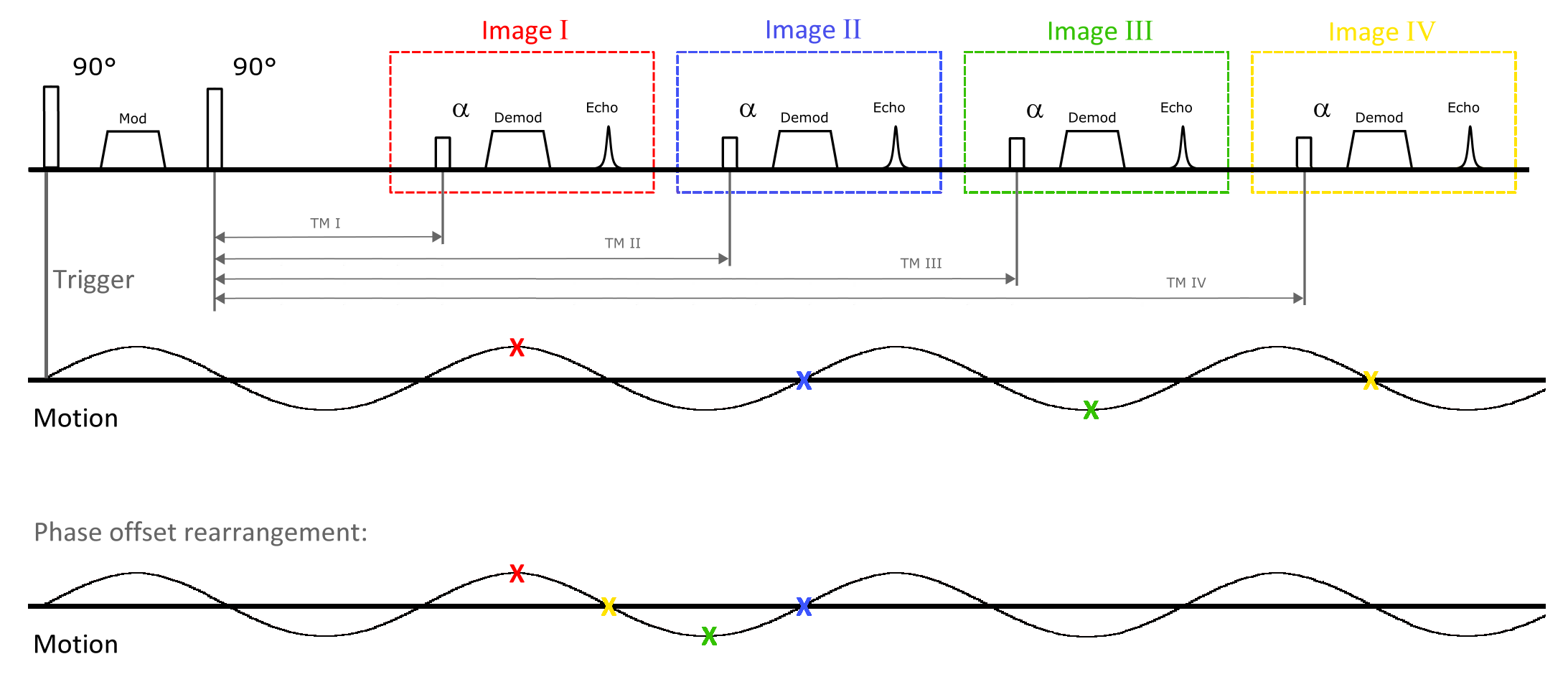
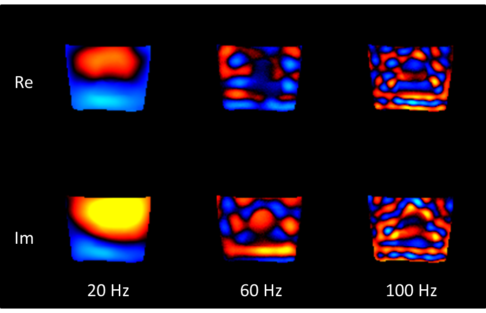
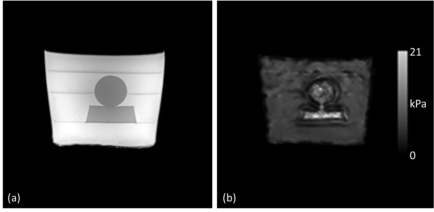
All experiments were performed with an agar phantom with two stiff inclusions on a 3T MRI scanner (MAGNETOM Prisma fit, Siemens Healthcare, Erlangen, Germany) using a 20ch head coil. The multi phase offset DENSE-MRE sequence was implemented with modulation and demodulation gradients in slice selection direction. Figure 1 illustrates the sequence diagram schematically. The following imaging parameters were used: matrix=128x128, FOV=300x300mm2, slice thickness=5mm, 15 slices, parallel imaging GRAPPA factor 2, α=14°, TE=9.5ms, mixing times=12ms+[0/12.5/25/37.5]ms. Thus 4 phase offset images were acquired consecutively at one run, by matching the mixing times to get equidistant sample time points of the vibration. The acquisition order of the sampling points was set in a way to keep the mixing times as short as possible. Rearrangement of the images on the time axis was performed afterwards. Thus the needed imaging time for all phase offsets of one slice during one TR was kept minimal in order to leave more time for interleaved multi-slice acquisition. Total acquisition time was 76s per frequency. Shear waves were excited by a piezoelectric driving unit which was connected to a head cradle for brain MRE. Continuous sine waves with 20Hz/60Hz/100Hz were used in a series of 3 measurements. Vibration synchronization was guaranteed by a trigger at each TR of 1000ms. To extract the wave propagation, either subtraction of the mean phase along time direction or picking the first harmonic after pixelwise Fourier transform of the 4 phase offset images from time to frequency domain was performed. To achieve estimates of the complex shear modulus, a multifrequency reconstruction (MDEV [2]) was used.Results
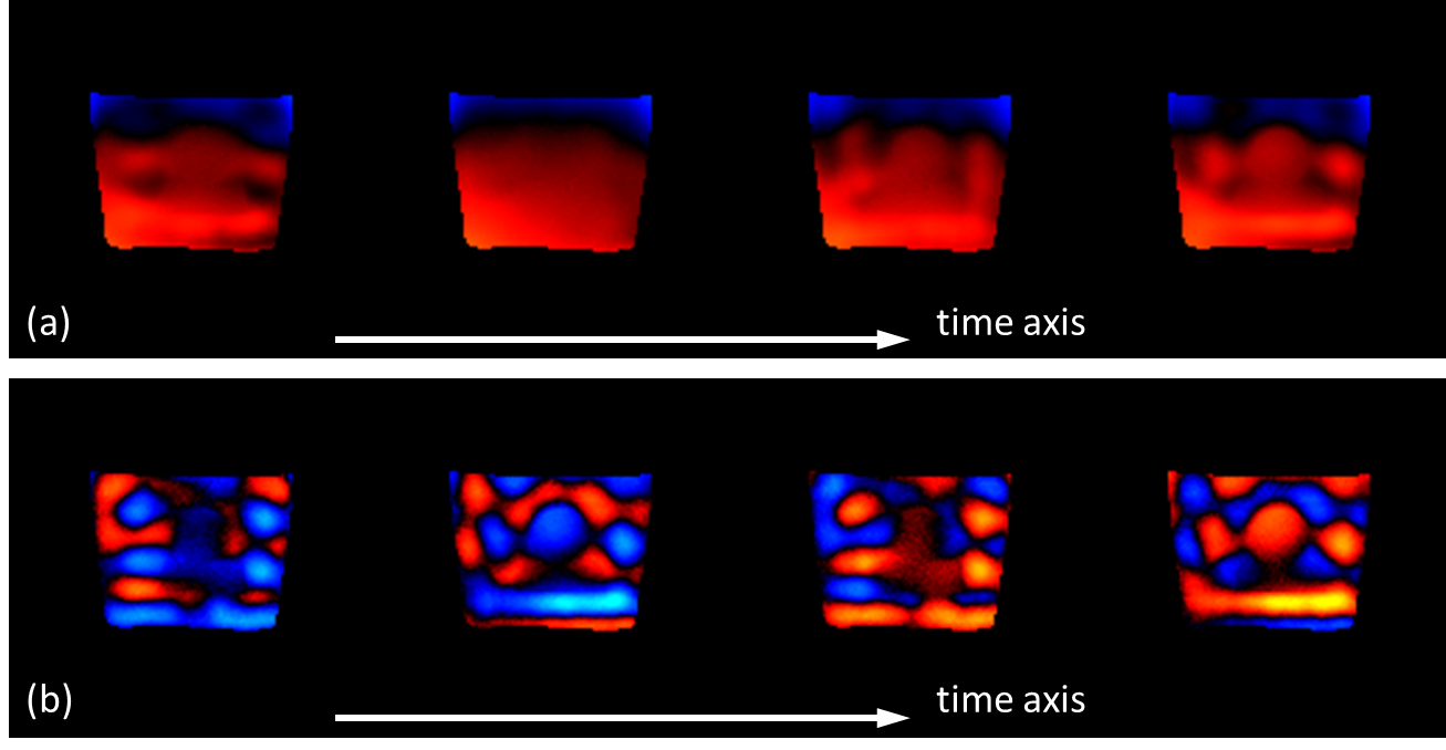
Using the proposed approach, 60 images (4 phase offsets, 15 slices) could be acquired within an acquisition time of 76s per frequency. After background phase removal, the artifact-free phase images clearly showed the propagation of the mechanical waves (figures 2 and 3). The magnitude of the estimated complex shear modulus clearly differentiated the two stiff inclusions from the background (figure 4).Discussion and conclusion
Our results indicate, that wave images in MRE can be acquired using DENSE together with a multi readout approach in a fast way. While the use of stimulated echoes comes along with an SNR reduction, it offers several advantages. The most significant ones are reduced susceptibility artifacts and shorter echo times. Both of them are becoming more relevant when moving towards lower vibration frequencies.Acknowledgements
The authors gratefully acknowledge Professor Ingolf Sack for providing the MDEV inversion algorithm.References
[1] Dittmann F et al., MRM 76:1116-1126 (2016)
[2] Hirsch S et al., MRM 71:267-277 (2014)
[3] Muthupillai R et al., Science 269:1854-1857 (1995)
[4] Robert B et al., MRM 62:1155-1163 (2009)
[5] Rump J et al., MRM 57:388-395 (2007)
[6] Aletras AH et al., J Magn Reson 137:247-252 (1999)
Figures