1329
Validation of the universal pulse concept at 7 Tesla on the Nova 8Tx/32Rx head coil with parallel transmit kT-point RF pulses1UNIRS, CEA/DRF/I2BM/Neurospin, Gif-sur-Yvette, France, 2Siemens Healthcare, Saint Denis, France, 3DZNE, Bonn, Germany
Synopsis
At ultra-high field, the use of parallel transmit (pTx) kT-point RF pulses can greatly improve the excitation uniformity of non-selective radiofrequency pulses but this approach generally requires additional pre-scans to map subject-specific transmit field sensitivities and compute optimal waveforms thereupon. Alternatively, quasi-optimal RF pulses can be obtained by replacing subject-specific field maps by a so-called field database resulting from the accumulation of field maps acquired in a small cohort. This concept is validated here at 7 Tesla on the Nova 8Tx/32Rx head coil with the implementation of a MP-RAGE protocol integrating universal kT-point pTx pulses.
Introduction
At ultra-high field (7 Tesla or higher), the use of parallel transmit (pTx) kT-point pulses1 can greatly improve the excitation uniformity of non-selective radiofrequency (RF) pulses. Usually, the optimal kT-point pulse is calculated from subject-specific transmit RF field (B1+) and static field offset maps (∆B0) measured in calibration scans through extensive optimization procedures, which unfortunately can limit the field of application of such approaches. Recently, it was suggested however that quasi-optimal kT-point pulses could be obtained for a given RF coil by optimizing the RF pulse coefficients upon a set of B1+ and ∆B0 measured separately on different subjects scanned with the same RF coil (field database)2-4. With this method, various pTx RF pulses in principle could be calculated offline to meet certain criteria (target flip angle, uniformity of excitation, specific absorption rate) and applied on any subject without any specific preparation, as it is usually done without pTx. In this work, we aim to validate this concept at 7T on the Nova 8Tx/32Rx head coil with the implementation of a MP-RAGE protocol integrating universal kT-point pTx pulses.Methods
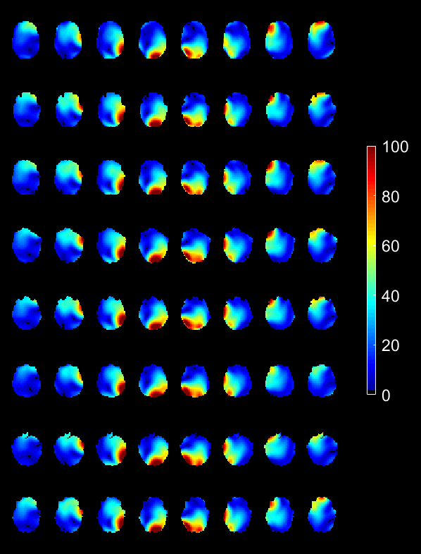
Experiments were performed on a pTX-enabled (Step 2.3) 7T Siemens scanner (Siemens Healthcare, Erlangen, Germany) equipped the Nova 8Tx/32Rx head coil (Nova medical Inc., Wilmington, USA) under local SAR supervision. The B1+ and ∆B0 mapping protocol consisted of a saturation prepared interferometric turbo-FLASH acquisition5 (5x5x5 mm3 voxels, matrix size 40x64x40, TR = 20 s, TA = 4’40’’) followed by a 3-echo 3D-GRE acquisition (2.5x2.5x2.5 mm3 voxels, matrix size 64x96x128, TR = 25 ms, TE = 5/6.5/8 ms, TA = 3’). With this protocol, a database of 8 different subject field maps was accumulated (see Fig. 1) and used to compute a so-called universal 7 kT non-selective small flip angle (FA) pulse (5°) under specific absorption rate (SAR) and power constraints, including RF coefficients and k-space trajectory optimization4. That pulse was then integrated in an MP-RAGE protocol (TR=2600 ms, TI = 1100 ms, TE = 3 ms, nominal FA = 5°, readout bandwidth 240 Hz, echo train length (ETL) 160, 1x1x1.1 mm3 voxels, 160x240x256 matrix size, GRAPPA acceleration factor 2, TA = 5’, adiabatic inversion) and was applied on five test subjects (not included in the field database). For comparison, the same MP-RAGE protocol was repeated this time with a standard non-selective 5° excitation where the transmit coil is driven in circularly polarized (CP) mode. Here the RF pulse amplitude was adjusted to achieve on average the prescribed FA in the central axial slice. For image comparison, the receive profile was estimated from a low-resolution proton density weighted GRE 3D acquisition (3x3x3 mm3 voxels, TR = 50 ms, TE = 2.3 ms, nominal FA = 5°, TA = 3’) and the appropriate intensity correction was applied to the MP-RAGE scans. The database field mapping protocol was finally applied to perform retrospectively pulse performance analysis.Results
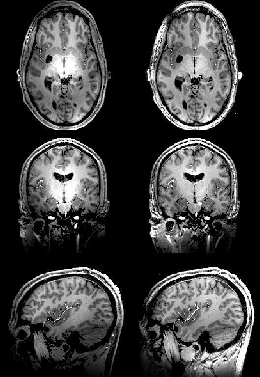
The FA-Normalized Root Mean Square Error (NRMSE) of the designed 5° pTx pulse evaluated across the 8 database subject was 9±1.5%. In comparison, the 5° CP pulse returned FA-NRMSEs of 27±1.7%. A qualitative comparison of the MP-RAGE images obtained on the first two test subjects is provided in Fig. 2 and 3 respectively. For the same visualization planes as displayed in Fig. 3, the simulated FA profiles for the inversion, the 5° CP and pTx universal pulses are provided in Fig. 4 for Subject 2.Discussion and conclusions
The CP versus universal pTx pulse comparison provided in Fig. 2 and 3 indicates that the universal pTx excitation optimized from a field database of 8 subjects allows enhancing the MP-RAGE signal and contrast in regions were the CP-mode typically lacks transmit efficiency. In addition, as shown in Fig. 4, the FA profile of the 5° pTx pulse shows only a mild residual non-uniformity, thus deemed acceptable for various UHF applications such as anatomical imaging. The remaining small inhomogeneity is due to an imperfect inversion of the adiabatic pulse at the bottom of the cerebellum, which should be substantially improved with the use of a universal inversion pulse4. To complete the validation of the universal pulse concept on the Nova 8Tx/32Rx head coil, additional test subjects will be included, the field database will be increased and the possibility to extend that concept to other standard MRI protocols such as multi-slice 2D gradient echo sequences will be studied.Acknowledgements
The research leading to these results has received funding from the European Research Council under the European Union's Seventh Framework Program (Proof of Concept UniPAT project).References
[1] Cloos et al. Magnetic Resonance in Medicine 2012, 67:72–80 [2] Jankiewicz et al. Journal of Magnetic Resonance 2010, 203:294–304 [3] Cloos et al. In Proceedings of the 19th Annual Meeting of ISMRM (2010), #3950 [4] Gras et al. Magnetic Resonance in Medicine 2010, DOI 10.1002/mrm.26148 [5] Fautz et al. In Proceedings of the 16th Annual Meeting of ISMRM (2008), p. 1247.Figures
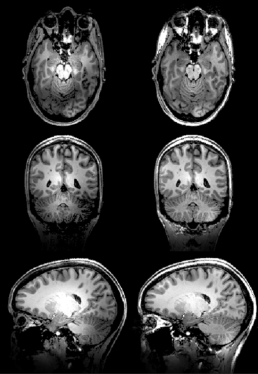
Receive profile corrected MP-RAGE images obtained in Subject 1. Left column: 5° CP excitation, right column: 5° pTx excitation. From top to bottom: representative axial, coronal and sagittal views through the brain. The occipital lobe and the right cerebellum which are under-excited in CP-mode appear well enhanced with the universal pTx pulse. Interestingly also, the bright center of the CP-mode is removed with the pTx excitation.

FA profile (deg) simulation based on the B1+ and ∆B0 maps of subject 2 for the 5° CP- and pTx-pulse and the 180° adiabatic pulse. The displayed axial, coronal and sagittal planes coincide with those visualized in Fig. 3. It is confirmed here that the CP-mode excitation is low in the occipital and temporal lobes and in the cerebellum. Additionally the bright center observed in the MP-RAGE images is clearly explained by the observed FA profile. The adiabatic pulse simulation on the other hand shows that the coil efficiency in the cerebellum region was not sufficient to invert the spins.