1319
Double-Spoke Slab-Selective Ramp Pulse Design for UHF TOF MR Angiography1CEA/DRF/I2BM/NeuroSpin/UNIRS, Gif-Sur-Yvette, France, 2Siemens Healthineers France, Saint-Denis, France, 3CEA/DRF/I2BM/NeuroSpin, Gif-Sur-Yvette, France, 4Laboratoires des Signaux et Systèmes, Université Paris-Saclay/CentraleSupélec/CNRS, Gif-sur-Yvette, France
Synopsis
Recently, the use of TOF with parallel transmission at Ultra High Field demonstrated an improvement in vessel-to-background contrast and spatial resolution. This study further investigates the in-vivo feasibility to correct for blood flow saturation effects in TOF slabs with a new double-spoke ramp pulse design optimization at 7T while mitigating in-plane TOF heterogeneities.
Purpose
The use of spoiled gradient-echo TOF at Ultra High Field (UHF) MRI recently demonstrated an improvement in vessel-to-background contrast and spatial resolution 1. However, radiofrequency (RF) field heterogeneities hinder the full visualization of the intravascular network 2. As previously reported 3, the method proposed hereby aims at compensating blood saturation effects by addressing a ramp of variable flip angles (FA) across a TOF slab while improving in-plane FA homogeneity by introducing a new 3D parallel transmission (pTX) pulse design optimization.Method
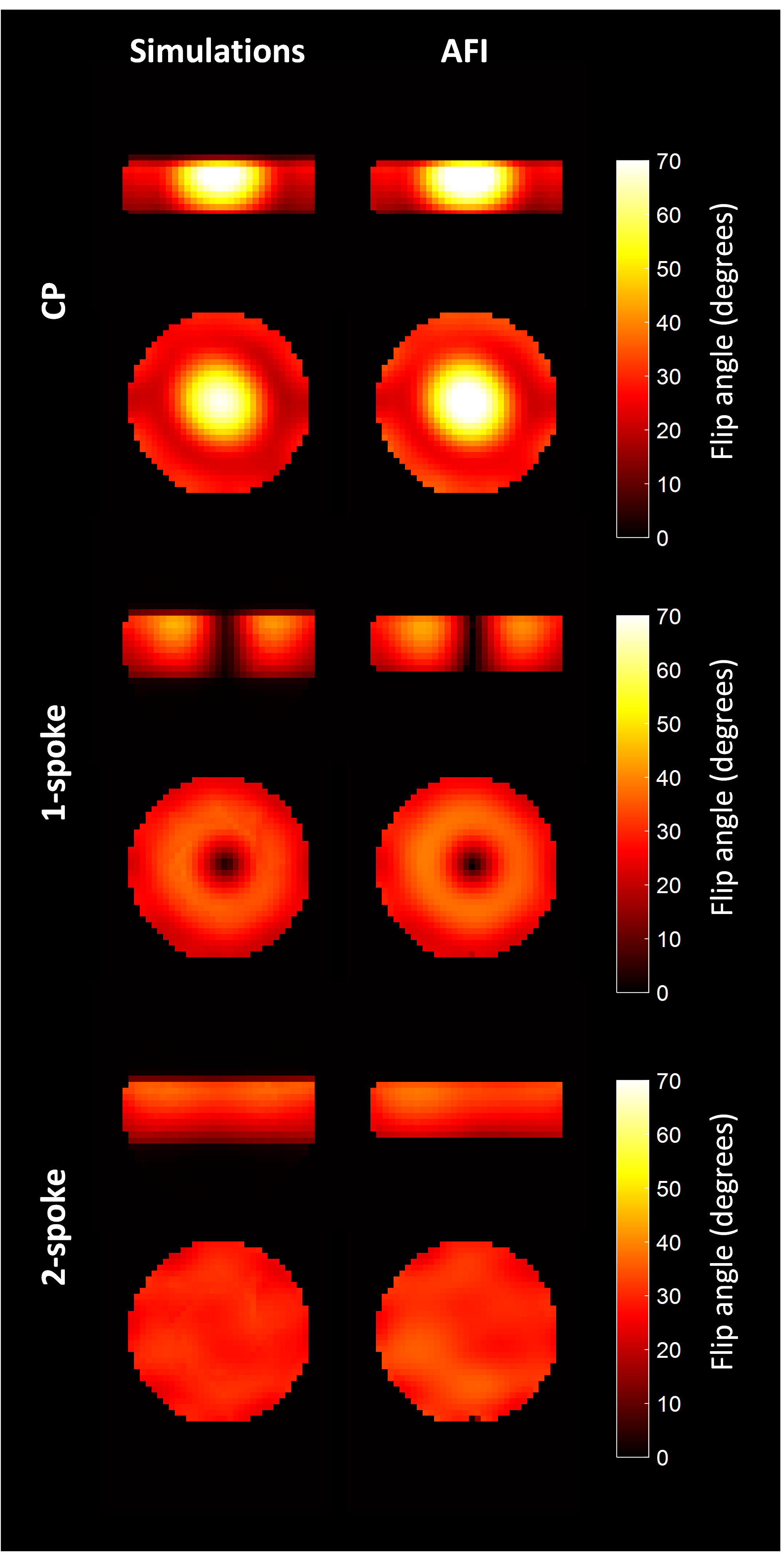
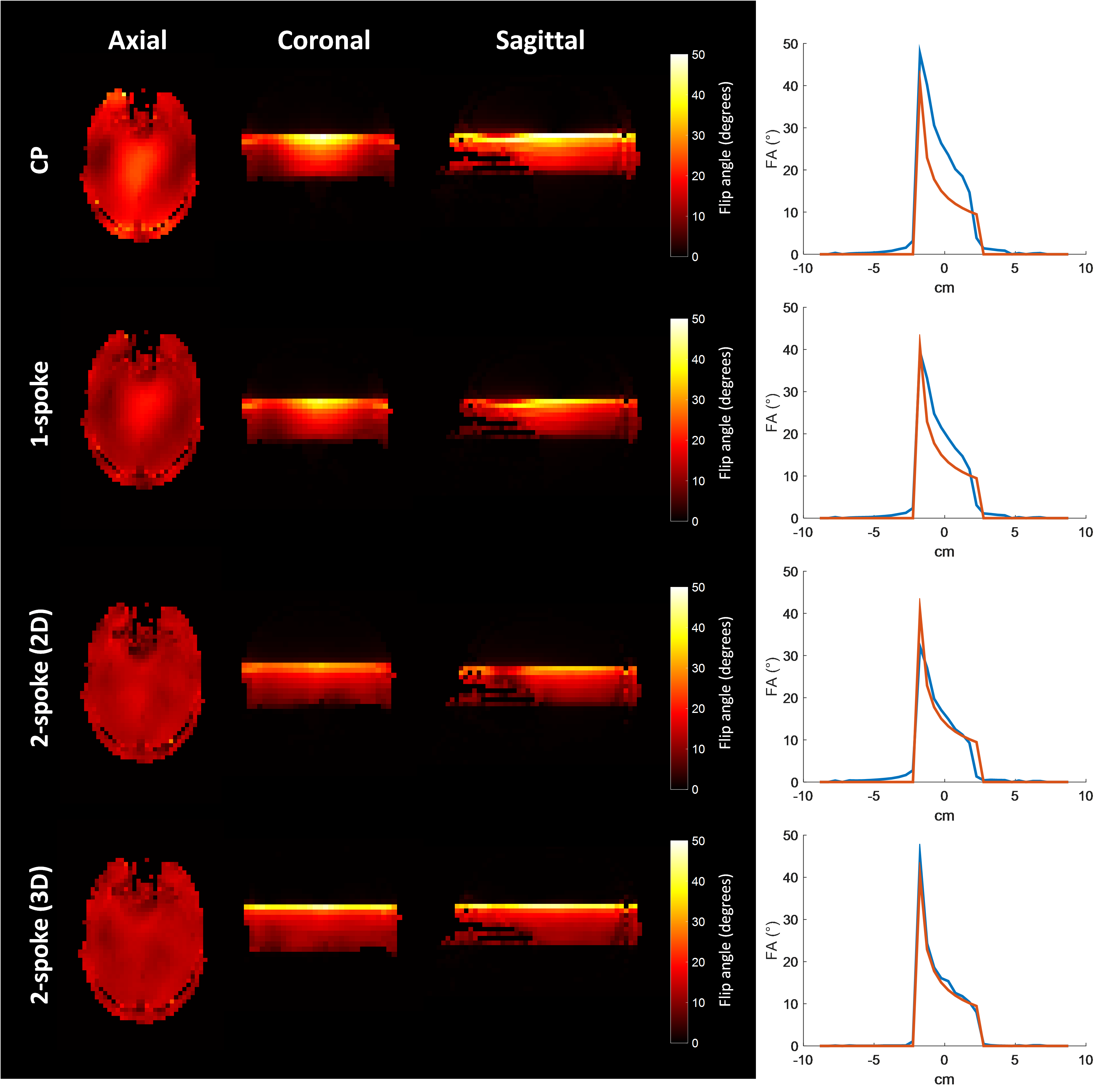
Slice selection in-plane B1+ heterogeneities can be compensated using kz “spokes” 4, especially in the pTX context: traditionally, complex weights of a predesigned RF waveform are optimized for each spoke and each channel by addressing a Magnitude Least Squares (MLS) two-dimensional problem. For TOF slab selection with a ramp of variable flip angles 5,6, this first approach is adopted here by predesigning a TONE or VUSE RF waveform. This was obtained by multiplying the frequency response of a TBW=9 sinc by the desired FA ramp, and by taking the inverse Fourier transform of the resulting profile. Then the 2D FA-target consists in a uniform slice in the middle of the slab. In a second approach, RF waveforms are a priori unknown and determined for every channel and spoke by addressing a full MLS three-dimensional problem targeting voxels in the slab with the desired FA profile, and all other voxels in the head with zero. Experiments were performed on a 7T scanner (MAGNETOM, Siemens AG, Healthineers, Erlangen, Germany) with an 8-Tx/Rx Rapid head array (RAPID Biomedical GmbH, Rimpar, Germany). B0 and B1-maps were acquired in a preliminary step to build the full k-space encoding matrix in the Small Tip Angle approximation. In the double-spoke experiments, the first spoke was placed in the center of the kx-ky plane while the second spoke was located at one inverse RF wavelength away from it (= 8m-1) 7. Both 2D and 3D optimizations were achieved with a variable exchange method initialization and Matlab’s (The Mathworks, Natick, MA, USA) active-set algorithm under strict constraints on local and global SAR, average power and peak amplitude. A 4.5-cm isocenter slab-selective TONE pulse was first tested on a doped spherical water phantom with a 20 to 40° FA ramp. The constrained pulse design was carried out for TR=20ms, which led to choose a pulse duration of 3ms to properly achieve the desired flip angle pattern. FA measurements in the slab were performed with an AFI sequence 8 to corroborate the FA pattern observed with Bloch equation simulations. Thus, the flip angle 3D distribution for the standard circularly polarized (CP) mode, static RF shimming (1-spoke) and 2-spokes were compared in terms of in-plane homogeneity and FA-ramp fidelity. An experiment was then conducted on a healthy informed volunteer using a spoiled GRE TOF pTX sequence with the following parameters: TR/TE = 20ms/7.8ms, resolution = 0.5x0.5x0.1mm3 , slice oversampling = 36.4% and TA=3.80min. The FA ramp was then changed into a non-linear VUSE profile between 9 and 40° to address low flow velocity (5 cm/s). Flow compensation was activated in slice and read directions to prevent blood motion artifacts.Results and Discussion
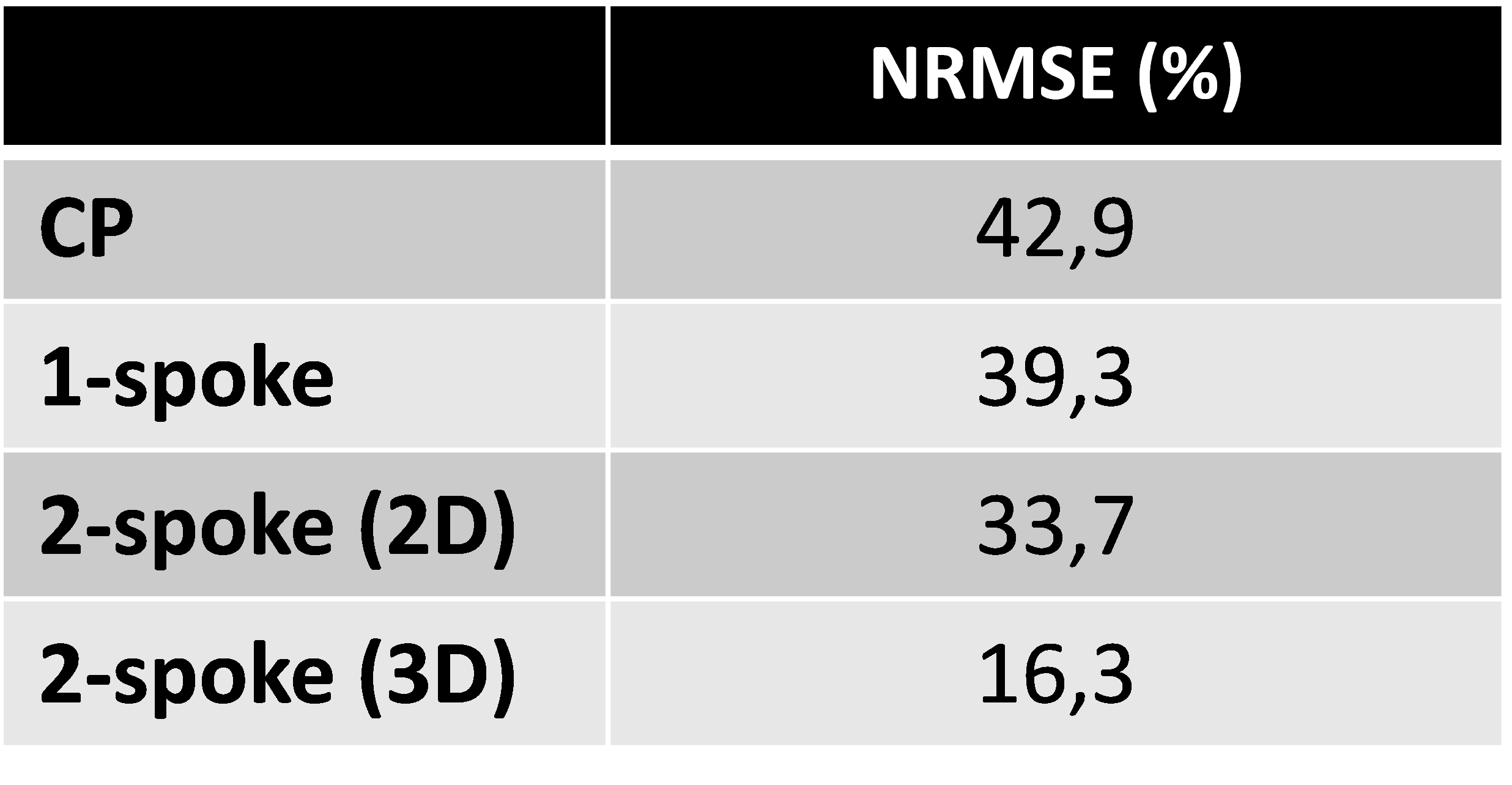
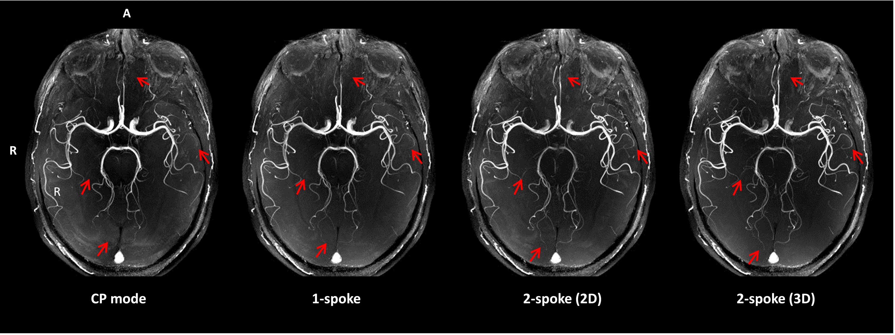
Based on simulations, the Normalized Root Mean Square Error (NRMSE) with respect to target FA’s in the slab was 37.1% for the CP mode, 19.2% for RF shimming, and 9.5% for the 2-spoke excitation (Figure 1). Simulated flip angle maps and AFI measurements were in good agreement. The RF shimming slightly improved the FA homogeneity compared to the CP mode despite the apparition of a hole in the center of the FA map. Nonetheless, this outcome was avoided using a second spoke. The 3D 2-spoke excitation significantly improved the flip angle in-plane homogeneity and ramp profile compared to the CP mode and more conventional pulse designs. In vivo results are presented in Figure 2 and Table 1 showing the outperforms of the 3D design to enhance details in the Maximal Intensity Projection (Figure 3).Conclusion
The visualization of the small arteries of the distal territories is significantly better using pTX. In addition, the 3D design distinctly improved the definition of the small vessels located in the posterior cerebral region compared to the traditional 2D design. Thus, this study demonstrates the ability to correct for blood flow saturation effects inside TOF rather large slabs in spite of intrinsic RF heterogeneities, with double-spoke pTX excitation tailored for a ramp profile at 7T.Acknowledgements
The authors wish to thank Chantal Ginisty for her precious help on MIP images post-treatment. We are grateful to Lucie Hertz Pannier, MD, PhD, for her advices and clinical evaluation of the in-vivo angiograms.References
1. Von Morze C, Xu D, Purcell DD, Hess CP, Mukherjee P, Saloner D, et al. Intracranial time-of-flight MR angiography at 7T with comparison to 3T. Journal of Magnetic Resonance Imaging. 2007 Oct;26(4):900–4.
2. Schmitter S, Wu X, Auerbach EJ, Adriany G, Pfeuffer J, Hamm M, et al. Seven-Tesla Time-of-Flight Angiography Using a 16-Channel Parallel Transmit System With Power-Constrained 3-dimensional Spoke Radiofrequency Pulse Design: Investigative Radiology. 2014 May;49(5):314–25.
3. Schmitter S, Wu X, Moeller S, Auerbach EJ, Adriany G, Van de Moortele P-F, and Ugurbil K, Slab selective pTX Multiband TOF Angiography at 7 Tesla, p. 542, proceedings ISMRM 2015, Toronto
4. Setsompop K, Wald LL, Alagappan V, Gagoski B, Hebrank F, Fontius U, et al. Parallel RF transmission with eight channels at 3 Tesla. Magnetic Resonance in Medicine. 2006 Nov;56(5):1163–71.
5. Atkinson D, Brant-Zawadzki M, Gillan G, Purdy D, Laub G. Improved MR angiography: magnetization transfer suppression with variable flip angle excitation and increased resolution. Radiology. 1994;190(3):890–4.
6. Priatna A, Paschal CB. Variablemangle uniform signal excitation (vuse) for threel dimensional time-of-flight MR angiography. Journal of Magnetic Resonance Imaging. 1995;5(4):421–7.
7. Amadon A. et al. Does the best distance beween 2 spokes match the inverse RF wavelength? p.2388, proceedings ISMRM 2015, Toronto.
8. Yarnykh VL. Actual flip-angle imaging in the pulsed steady state: A method for rapid three-dimensional mapping of the transmitted radiofrequency field. Magnetic Resonance in Medicine. 2007 Jan;57(1):192–200.
Figures