1318
Sheared 2DRF Excitation for Improved Off-resonance Robustness in Reduced FOV Imaging1Department of Electrical and Electronics Engineering, Bilkent University, Ankara, Turkey, 2National Magnetic Resonance Research Center (UMRAM), Bilkent University, Ankara, Turkey, 3Global MR Applications & Workflow, GE Healthcare, Menlo Park, CA, United States
Synopsis
Reduced field-of-view (FOV) using two-dimensional spatially selective radio frequency (2DRF) excitation has been widely used for targeted, high-resolution diffusion weighted imaging (DWI). This work proposes a sheared 2DRF excitation scheme to rapidly and efficiently cover the excitation k-space. This approach not only enables extended slice coverage and preserves fat suppression capabilities, but also significantly improves the robustness against off-resonance-induced signal losses of 2DRF pulses.
Purpose
To improve off-resonance robustness of two-dimensional radiofrequency (2DRF) excitation pulses for high-resolution, extended-slice-coverage reduced field-of-view (FOV) imaging, while providing robust fat suppression.Introduction
Reduced-FOV diffusion weighted imaging (DWI) has been widely used for high-resolution targeted imaging [1-8]. The reduced-FOV approach using 2DRF pulses excites a small phase-FOV extent, allowing high-resolution imaging with fewer phase-encode (PE) lines. In this technique, the 2DRF pulse has to be sufficiently long (16-20 ms) to allow imaging of multiple slices [8]. Previously, rotation of the excitation k-space was proposed to overcome the slice-coverage limitations [6,7], but this process further prolonged the 2DRF pulse. An important side effect of long RF pulses is sensitivity to off-resonance-induced signal losses. This work proposes a sheared 2DRF excitation scheme to rapidly and efficiently cover the excitation k-space. The proposed design significantly improves the robustness against off-resonance-induced signal losses, while preserving fat suppression capabilities and removing slice coverage limitations.Methods
2DRF Pulse Design
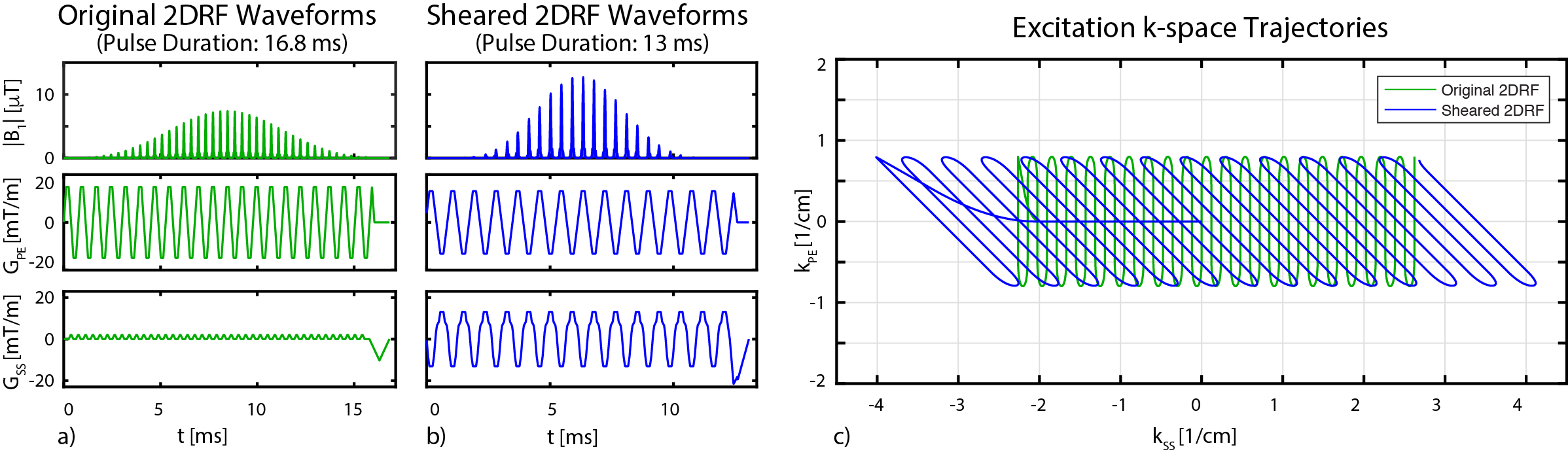
The 2DRF pulse designs were implemented in MATLAB. First, an original 2DRF pulse with a regular echo-planar trajectory was designed for FOVPE = 4cm, with slice thickness Δz = 5mm, time-bandwidth product (TBW) in the PE and slice-select (SS) directions TBWPE = 6 and TBWSS = 3.1, and 16.8 ms pulse duration. This 2DRF pulse allowed imaging a maximum of 16 slices without experiencing partial saturation effects due to sidelobes along the SS direction. Next, a sheared trajectory that covers the same excitation k-space using fewer lines with 45°-degree shearing angle was created. The resulting sheared 2DRF pulse had a significantly shorter pulse duration of 13 ms. In principle, the sheared 2DRF and the previously presented tilted 2DRF [7] exhibit similar 2D excitation profiles, but a sheared design allows a more efficient coverage of the excitation k-space, and is less demanding of the gradient performance.
Off-Resonance Robustness
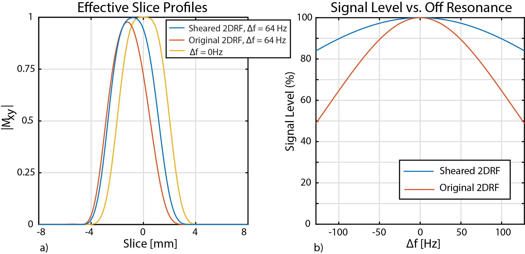
The off-resonance robustness of the original and sheared 2DRF pulses were compared via Bloch simulations. A 180° RF pulse with 3.2 ms duration was used as the refocusing RF pulse. The combination of 2DRF and 180° RF pulses provide a fat suppression capability to the reduced-FOV imaging technique [1,7,8]. The effective slice profiles were simulated for ±128 Hz off-resonance (i.e., ±1 ppm at 3T), and the overall signal levels were computed by integrating the simulated 2D excitation profiles along the slice direction.
Simulations on Brain Phantom
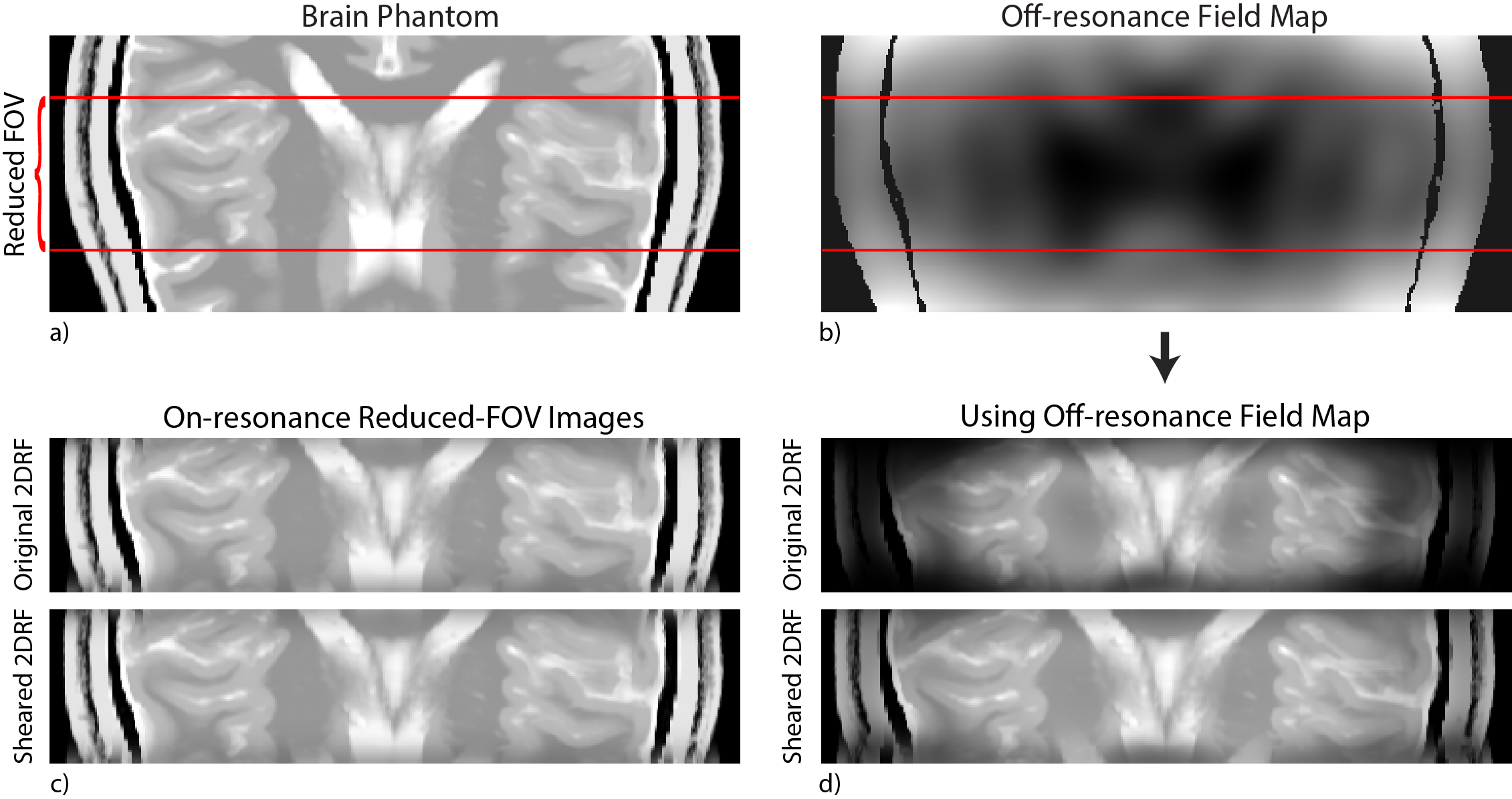
A T2-weighted digital brain phantom was generated from a fuzzy brain model [9], and interpolated to a fine 3D grid (0.5x0.5x0.25 mm3 voxel size), as shown in Fig. 4a. A realistic off-resonance field map was generated based on the anatomy of the phantom (Fig. 4b), where the off-resonance within the reduced-FOV imaging section was 97±82 Hz (mean±std). The resulting reduced-FOV images were generated via Bloch simulations for both the sheared and the original 2DRF pulses.
Results and Discussion
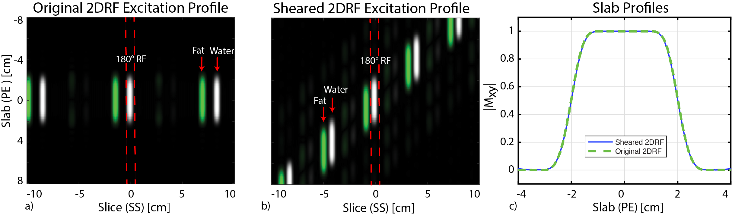
The waveforms and excitation k-space trajectories of the original and sheared 2DRF pulses are shown in Fig. 1. The corresponding 2D excitation profiles are given in Fig. 2, where the excitation sidelobes are pushed outside the imaging section in the sheared design, while maintaining the sharpness in slab profile (Fig. 2c). As in the original design, the sheared 2DRF pulse still allows fat suppression via a subsequent 180° RF pulse (Fig. 2b).
The effective slice profiles for on-resonance and 64 Hz off-resonance cases are shown in Fig. 3a, and the overall signal levels for ±128 Hz off-resonance are plotted in Fig. 3b. The underlying cause for the off-resonance sensitivity is the mismatch between the pulse durations of the 2DRF and 180° RF pulses. As a result, the excitation and refocusing profiles overlap only partially, thinning the effective slice profiles. Since the sheared 2DRF pulse has a shorter duration, this problem is significantly alleviated. Quantitatively, while the overall signal level for 128 Hz off-resonance drops to 49% for the original 2DRF case, the signal level remains at 84% for the sheared 2DRF case. Ratio-wise, this corresponds to a remarkable 71% SNR improvement at 128-Hz off-resonance.
The brain phantom simulations show the images for fully on-resonance case, as well as for the off-resonance field map given in Fig. 4b. Comparing Fig. 4c and 4d, the sheared 2DRF technique demonstrates significant improvement in robustness against off-resonance-induced signal losses.
Conclusion
The proposed sheared 2DRF technique significantly improves the off-resonance robustness of the reduced-FOV imaging technique, while removing slice coverage problems and preserving fat suppression capabilities. This technique will be especially useful for high-resolution reduced-FOV imaging of difficult anatomies, such as the spinal cord and the breast.Acknowledgements
This work was supported by the European Commission through an FP7 Marie Curie Career Integration Grant (PCIG13-GA-2013-618834), by the Turkish Academy of Sciences through TUBA-GEBIP 2015 program, and by the BAGEP Award of the Science Academy.References
1. Saritas EU, Cunningham CH, Lee JH, Han ET, Nishimura DG. DWI of the spinal cord with reduced FOV single-shot EPI. Magn Reson Med 2008;60:468–473.
2. Finsterbusch J. High-resolution diffusion tensor imaging with inner field-of-view EPI. J Magn Reson Imaging 2009;29:987–993.
3. Wheeler-Kingshott CA, Parker GJ, Symms MR, Hickman SJ, Tofts PS, Miller DH, Barker GJ. ADC mapping of the human optic nerve: increased resolution, coverage, and reliability with CSF-suppressed ZOOM-EPI. Magn Reson Med 2002;47:24–31.
4. Wilm BJ, Svensson J, Henning A, Pruessmann KP, Boesiger P, Kollias SS. Reduced field-of-view MRI using outer volume suppression for spinal cord diffusion imaging. Magn Reson Med 2007;57:625–630.
5. Heidemann RM, Anwander A, Feiweier T, Knösche TR, Turner R. Turner. k-space and q-space: combining ultra-high spatial and angular resolution in diffusion imaging using ZOOPPA at 7 T. NeuroImage 2012;60:967–978.
6. Finsterbusch J. Improving the performance of diffusion-weighted inner field-of-view echo-planar imaging based on 2D-selective radiofrequency excitations by tilting the excitation plane. J Magn Reson Imaging 2012;35:984–992.
7. Banerjee S, Nishimura DG, Shankaranarayanan A, Saritas EU. Reduced field-of-view DWI with robust fat suppression and unrestricted slice coverage using tilted 2D RF excitation. Magn Reson Med. DOI: 10.1002/mrm.26405.
8. Saritas EU, Shankaranarayanan A, Zaharchuk G, Nishimura DG. Reduced-FOV single-shot diffusion-weighted EPI: extended slice coverage with tailored RF pulse design. In Proceedings of the 19th Annual Meeting of ISMRM, Montreal, Quebec, Canada, 2011, p. 1953.
9. Cocosco CA, Kollokian V, Kwan RKS, Pike GB, Evans AC. Brainweb: Online interface to a 3D MRI simulated brain database. NeuroImage 1997; 5:425.
Figures